Thursday, December 3 | 11:30 a.m.-11:40 a.m. | SSQ10-07 | Room S402AB
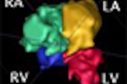
A team from Stanford University will share a new method for processing and interpreting 4D flow MRI studies in this Thursday morning scientific session.While 4D flow MRI (time-resolved, 3D phase-contrast MRI) is a promising technique for dynamic characterization of blood flow, its clinical utility has been limited to date by a lack of computational software tools for reading and interpreting the data, said presenter Dr. Albert Hsiao, Ph.D., of Stanford University in Stanford, CA.
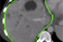
These tools are needed to distinguish intravascular signals from lung, to segment vascular structures, to calculate physiologic parameters, and to visualize flow in a meaningful way for diagnostic interpretation, according to the researchers.
In his talk, Hsiao will describe how the combination of semiautomated vessel segmentation with intelligent imaging planes can reliably calculate clinically relevant quantities from 4D flow MRI data.
"With the right computational tools, 4D phase-contrast MRI is feasible for basic qualitative and quantitative analysis of flow dynamics," Hsiao said. "A lot more needs to be done to show how reliable it is compared to conventional 2D phase-contrast MRI and Doppler echocardiography, but I think it has promise."


