Thursday, December 3 | 11:50 a.m.-12:00 p.m. | SSQ10-09 | Room S402AB
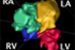
A new automatic segmentation method can outperform traditional segmentation methods used in delayed gadolinium-enhanced MRI for assessing myocardial viability.A joint research effort between Fraunhofer MEVIS Institute for Medical Image Computing in Bremen, Germany, and the Hospital Tübingen in Germany compared their automatic segmentation method with manual delineation of reference regions by clinical experts and the widely-used 3SD threshold segmentation method in 20 MRI studies.
The automatic method had higher correlation with the manual delineation results than 3SD. Presenter Anja Hennemuth from Fraunhofer MEVIS will share more details on the method in her Thursday talk.
"In the long term, we hope to achieve a reliable automatic classification of the myocardium into healthy regions, myocardium at risk, and already-necrotic tissue to allow an assessment of the achievable benefit through reperfusion therapy," she said.


