Tuesday, December 2 | 10:30 a.m.-10:40 a.m. | SSG07-01 | Room S402AB
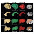
A team from Johns Hopkins University will explore the potential of texture analysis to assist in the classification of renal lesions on CT scans.Texture analysis is a method designed to quantify heterogeneity within a region of interest on an imaging study. While other research groups have mostly investigated texture analysis for assessing tumor treatment response, the Johns Hopkins team sought to apply the technique to provide quantitative information to help predict the histology of a tumor seen on a CT scan, said presenter Dr. Siva Raman.
Predicting the histology of renal masses on CT is currently performed by techniques such as visually assessing tumors and measuring Hounsfield attenuation values; however, the researchers wanted to see if they could create predictive models based on texture data that could differentiate several basic types of renal masses, including clear-cell renal cell carcinoma, papillary renal cell carcinoma, and oncocytoma, Raman said.
In testing, the models were highly accurate, even for lesion types such as clear-cell renal cell carcinoma and oncocytomas that can't be readily distinguished visually, he said. While acknowledging that this was just a small pilot study, Raman said the results suggest there is long-term potential for quantitative modeling to aid radiologists in formulating final diagnoses.
"Clearly, there is helpful data hidden in the CT scan that we as radiologists cannot appreciate just by visually looking at the study, and the key is to come up with methods to obtain that data and then create modeling techniques that can utilize that data to help formulate a diagnosis," Raman told AuntMinnie.com. "It is not far-fetched to think that someday the radiologist will be able to draw a [region of interest] around a tumor, derive a few statistical parameters (such as from texture analysis), plug those numbers into a statistical model, and have a better chance of correctly predicting a tumor's histology than by just visually examining the tumor."

