Tuesday, December 2 | 11:00 a.m.-11:10 a.m. | SSG07-04 | Room S402AB
In this presentation, researchers from Weill Cornell Medical College will show how quantification of a donor's kidney volume offers insight into whether a kidney transplant will succeed.Transplantation medicine is one of the few areas of medicine in which operations occur in healthy people; kidney donors voluntarily undergo surgery to have one of their healthy kidneys removed and given to a needy recipient, said senior author Dr. Krishna Juluru.
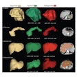
Prior to surgery, many donors receive a CT angiogram, followed by 3D reconstructions to characterize the vasculature to the kidneys, Juluru said. Those same exams and advanced visualization tools were used by the researchers to quantify the donor's kidney size, both its cortex and entire volume.
The group then worked with colleagues from the transplantation nephrology department to determine if these additional quantitative parameters offered any value for predicting the one- and two-year success of the renal transplant.
"We found that after adjusting for a variety of other transplantation variables, the ratio of the size of the donated kidney to the size of the recipient is an independent marker of transplantation success at one and two years," he said.
While acknowledging that there has been similar work in this area, Juluru noted that the kidney size measurements were performed after brief training by two college students with limited medical background. These measurements were highly reproducible, he said.
"This evidence of reproducibility is important for researchers such as myself and groups such as RSNA's Quantitative Imaging Biomarkers Alliance who are interested in moving radiology from a qualitative to a quantitative discipline," Juluru told AuntMinnie.com. "Furthermore, the study adds to a growing body of literature on how imaging can be used as a biomarker."
Learn more by checking out this presentation, which will be given by first author Dr. Jessica Rotman.


