Adding bone-suppression functions to digital chest x-ray exams can assist doctors with the detection of subtle pulmonary lesions, according to a research group in Japan.
Researchers at the University of Occupational and Environmental Health in Fukuoka found that suppressing bone artifacts on temporal subtraction (TS) digital chest radiographs significantly improved visual evaluation of pulmonary lesions. The study is the first to demonstrate the clinical usefulness of the approach in patients, the authors wrote.
"This study demonstrated that [bone suppression]-TS processing can reduce rib and clavicle artifacts, and this method can assist in the detection of subtle pulmonary lesions in digital chest radiography," noted lead author Takeshi Takaki, PhD, and colleagues. The study was published online January 16 in Heylion.
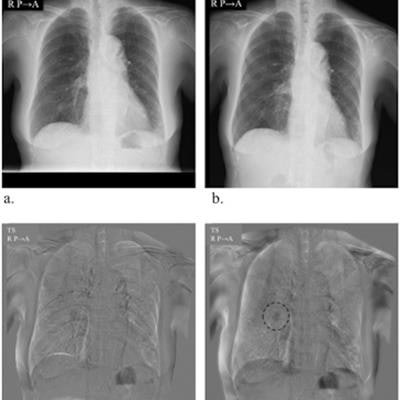
TS during digital chest x-ray image processing is an emerging technique that has shown promise for improving the sensitivity of nodule detection. The method subtracts normal structures such as the ribs or clavicle from a current chest image using a previous corresponding image from the same patient, and thus reveals potentially abnormal findings in overlapping areas.
However, artifact reduction is also necessary in TS processing because differences in patient positioning at the time of imaging can cause bone-related artifacts, possibly resulting in false-positive findings, according to the authors.
In previous studies, bone suppression has improved radiologists' detection of lung nodules on digital chest x-rays. In this reader study, the researchers examined differences in detection between conventional TS (C-TS) and TS using bone suppression (BS-TS).
Five radiologists were recruited to evaluate images processed using both the C-TS and BS-TS techniques. Images were selected from 31 patients with lung disease, including 19 with primary lung cancer, eight with lung metastases, and four with pneumonia. A majority of patients' lesions were classed as subtle, very subtle, or extremely subtle on prior CT scans.
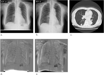 Images used for temporal subtraction (TS). (a) Previous radiograph. (b) Current radiograph. (c) CT image. (d) Conventional TS image. (e) Improved TS image acquired with bone suppression (BS) processing. The rib artifacts are reduced with BS-TS, and nodule areas are easily recognized. Image and caption courtesy of Heylion through CC BY 4.0.
All chest radiographs were acquired using a flat-panel detector (AeroDR 1717HQ, Konica Minolta) and processed using a TS imaging workstation with updated software that included bone-suppression processing functions.
The performance -- as assessed by figure-of-merit values -- of all radiologists increased significantly using the bone-suppression method, from 0.619 (conventional) to 0.696 (p = 0.032). The average sensitivity for detecting pulmonary lesions improved from 67.9% to 75.4%, and the average number of false positives per case decreased from 0.336 to 0.252 using bone suppression temporal subtraction.
"The overall observers' ability to detect new lesions increased significantly with the use of BS-TS; this method allowed the enhancement of changes in the density of faint lesions, providing higher image quality than C-TS," the authors stated.
Ultimately, the authors noted that lung anatomy is complex and that the range of diseases possibly present on a chest radiograph is extensive. With further development, the BS-TS technique could become a useful tool even for experienced radiologists, they wrote.
"Further observer performance studies including experienced radiologists with a larger number of cases are likely necessary to confirm the clinical usefulness of this computerized method," Takaki and colleagues concluded.


