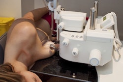
The level of a woman's background parenchymal enhancement (BPE) on MRI impacts the modality's performance for detecting breast cancer, according to research presented at the recent RSNA meeting in Chicago.
The findings underscore the need to keep BPE levels in mind when using MRI to screen or diagnose breast cancer -- and adjust protocols accordingly, said presenter Dr. Sarah Eskreis-Winkler, PhD, of Memorial Sloan Kettering Cancer Center in New York City. BPE is the enhancement of normal fibroglandular tissue on contrast-enhanced dynamic breast MRI.
"According to several studies, increased BPE is associated with higher biopsy rates and lower specificity, [suggesting] that more work is needed to optimize breast MRI protocols for patients with high BPE," she said.
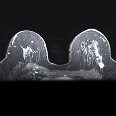
MRI is the best available tool for breast cancer detection, finding 15 cancers per 1,000 women compared with the seven or eight per 1,000 women found with mammography plus ultrasound, Eskreis-Winkler said. But background parenchymal enhancement is considered a risk factor for breast cancer, as it can obfuscate the disease and is known to complicate the interpretation of breast MRI. High background parenchymal enhancement is associated with making interpreting breast MRI exams more difficult, as well as higher biopsy rates and lower specificity.
"Despite the logical concern that high BPE might mask cancers and decrease cancer detection, no study to date has found a statistically significant decrease in cancer detection rate for high versus low BPE exams," the team wrote in its abstract about the research. "To further investigate this, we compared the diagnostic performance of breast MRI in patients with high versus low BPE."
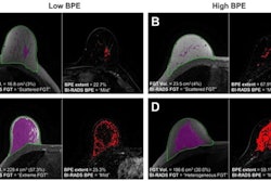
Eskreis-Winkler's group assessed breast MRI's performance by BPE levels (high levels, categorized as "marked" or "moderate," and low levels, categorized as "mild" or "minimal"). The team also evaluated two different breast MRI protocols: a standard, high spatial resolution protocol, and a simultaneous high temporal/high spatial resolution (HTHS) protocol.
The research included 2,933 MRI exams performed between January and December 2016, 23.3% of which were baseline studies and 76.7% of which had prior MRI exams available for consideration. Of the total number of exams, 22.7% were categorized as having high BPE and 77.2% as having low BPE.
Eskreis-Winkler and colleagues found that, overall, higher background parenchymal enhancement decreased cancer detection rates and positive predictive values, increased interval cancer rates, and lowered sensitivity and specificity.
| Performance of breast MRI depending on BPE level | ||
| Measure | Low BPE | High BPE |
| Cancer detection rate | 0.22% | 0.12% |
| Positive predictive value | 29.7% | 9.3% |
| Interval cancer rate | 0.1% | 0.6% |
| Sensitivity | 93% | 66.7% |
| Specificity | 94.5% | 88.1% |
But they also found that the high temporal/high spatial resolution protocol outperformed the standard one in women with high BPE, finding 24 cancers per 1,000 women, compared with eight out of 1,000 identified with the standard protocols, and increasing the positive predictive value from 6% to 16% compared with standard breast MRI, Eskreis-Winkler said.
It's clear that women's background parenchymal enhancement levels should be taken into account and breast MRI protocols adjusted accordingly, she noted.
"Breast MRI protocols with high spatiotemporal resolution may be warranted in high BPE patients to visualize lesions at an earlier post-contrast timepoint, avoiding BPE-induced lesion obscuration," Eskreis-Winkler concluded.