CHICAGO -- CT and ultrasound have elevated importance for bomb blast care in areas of conflict, according to research presented December 3 at RSNA 2024.
Zahra Rahmatullah, a final-year medical student from Aga Khan University Hospital in Karachi, Pakistan, presented her team’s findings on imaging trends for abdominal and pelvic injuries for these patients, with CT and ultrasound being the modalities most used.
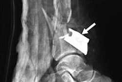
“Imaging is pivotal. Many of these patients undergo multiple different scans,” Rahmatullah told AuntMinnie.com. “CT is the best method to check for any abdominal penetrations, especially when it comes to shrapnel and shrapnel-related injuries.”
Pakistan has been an area of conflict for years. Previous reports indicate that 63 bomb blasts took place in Karachi from 2000 to 2007. And between 2007 and 2011, the city experienced 46 explosions. Rahmatullah illustrated this with news headlines on such attacks whether by groups or suicide bombings.
"Tragically, for the people of Pakistan, headlines like these are all too familiar," she said.
Bombings that involve explosives packed with metal fragments can alter the patterns and severity of injuries. This means imaging is necessary to determine treatment strategies, though MRI’s use is limited in these cases. Rahmatullah said that patients are referred for advanced care if smaller healthcare facilities do not have the equipment or personnel to manage these events.
The researchers uncovered patterns and implications of abdominal and pelvic injuries in bomb blast victims through radiological and clinical profiling.
The study included 160 patients who presented between 2004 and 2024 for bomb blast injuries and had their injuries confirmed via imaging. The team retrieved data from electronic health records. More men were included in the study than women at a 15:1 ratio.
The team considered about 43% of injuries to be secondary and another 34% to be a combination of secondary and tertiary injuries. Nearly 30% of the injuries could not be characterized.
CT exams made up the most common imaging method at 42% on first investigation, with ultrasound being next at 35%. Digital x-ray, meanwhile, was third at 17%. Fluoroscopy, angiography, and nuclear scans all came in at less than 5%.
For second investigations though, ultrasound led the pack at 34%, followed by CT at 26%. X-ray’s use in second investigations increased to 28%.
For injury distribution, “normal” cases constituted most cases on first and second investigations. Hemoperitoneum and pneumoperitoneum cases were the next most common cases on first investigations.
Finally, the team observed that low-grade injuries that did not require surgical management were more common than high-grade injuries for liver, spleen, and kidney injuries. Patients who underwent abdominal and/or pelvic surgery spent an average of 11.81 days in the hospital, while those who did not stayed for 7.96 days on average.
Rahmatullah highlighted that these findings can aid in care strategies for areas with high conflict, including those with limited resources.
“This could be pivotal as in low- to middle-income countries, resources are limited, CTs are expensive, and patients may be paying out of pocket,” she told AuntMinnie.com. “Ultrasound and x-rays … proved to be just as useful for us. We think they can be cost-effective alternatives to CT, especially for second investigations.”
For full coverage of RSNA 2024, visit our RADCast.
