Radiol Clin North Am 1994 Jul;32(4):759-774
Helical computed tomography of the thorax. Clinical applications.
Naidich DP
Department of Radiology, New York University Medical Center, Bellevue Hospital, New York.
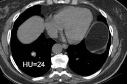
Intuitively, any technique that minimizes the effects of respiratory motion, eliminates misregistration between scans, minimizes intravenous contrast requirements, and allows high quality multiplanar and 3-D image reconstruction is likely to have a tremendous impact on conventional notions concerning routine thoracic CT. Helical scanning is already of proved efficacy for vascular and airway imaging as well as for identifying and characterizing pulmonary nodules. It may be anticipated that the indications for the use of helical imaging will continue to expand. Of particular interest is the ongoing development of reconstruction algorithms that allow high-quality images to be obtained with rapid table incrementation while simultaneously reducing radiation exposure. Given the intrinsically high contrast of structures within the thorax coupled with the disadvantages that result from respiratory motion, it is not unreasonable to conclude that within the near future volumetric techniques will be the standard for nearly all CT applications within the thorax.
PMID: 8022979, MUID: 94294604
