Dear AuntMinnie Member,
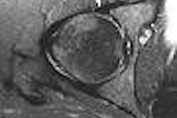
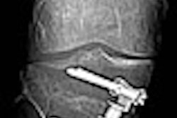
MRI may be able to improve its standing as a modality for imaging labral tears, and for selecting patients most suited for arthroscopy, thanks to a technique developed by U.S. researchers.
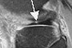
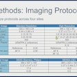
MRI has historically demonstrated low accuracy for diagnosing labral tears. But researchers at Duke University in Durham, NC, have improved its performance through the use of contrast media and a small field-of-view. That's according to an article we're featuring this week in our Orthopedic Imaging Digital Community by staff writer Shalmali Pal.
Using 1.5-tesla MRI and the technique, they were able to improve MRI's sensitivity to 92%. Find out how they did it by clicking here.
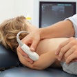
Another orthopedic imaging story features researchers from Thomas Jefferson University in Philadelphia, who share their technique for ultrasound-guided percutaneous needle tenotomy as an alternative to surgery for patients with tennis elbow.
They've found that the technique is a less-invasive alternative to a surgical method in which the tip of a scalpel is used to release the tendon fibers and stimulate new blood vessels at the site of the tendinitis. Read all about it by clicking here.
For more news from the world of orthopedic imaging, check out the Orthopedic Imaging Digital Community at orthopedic.auntminnie.com.

