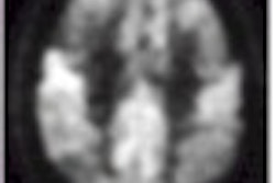
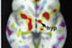
SPECT brain scans of children with attention deficit/hyperactivity disorder (ADHD) typically show evidence of infraorbital prefrontal deactivation, says a researcher from Columbus Children’s Hospital in Ohio. These areas of the brain modulate executive functioning such as organization, impulse control, and judgment.
Dr. Larry Binkovitz reviewed the spectrum of nuclear brain scan abnormalities in children with ADHD at the November RSNA conference in Chicago. Binkovitz’s co-authors included Dr. Daniel Amen, founder of the Amen Clinic for Behavioral Medicine in Fairfield, CA.
"Based on alterations of cerebral blood flow depicted with nuclear brain SPECT imaging, Amen described five patterns for brain scan abnormalities found in ADHD. All five show decreased activity in the infraorbital and prefrontal cortices," Binkovitz said.
"Four subtypes also show findings thought to reflect abnormalities in neural circuits separate from the prefrontal cortex," Binkovitz added. These subtypes are prefrontal, cingulate, limbic, temporal, and ring-of-fire.
The study group consisted of 100 patients, ages 7-17, who had been referred to an outpatient behavioral clinic for a brain scan. Most of the patients -- 51 males and 22 females -- had been diagnosed with attention deficit disorder (ADD), while only a few were diagnosed with the additional hyperactivity disorder.
"The majority of these patients, not having been diagnosed with ADHD, had not been treated with previous drug treatments," Binkovitz said. "Psychiatric comorbidity is very frequent in ADHD, as it was in this group. Of the 73 patients, 57 had additional psychiatric diagnoses," such as depression, bipolar disorder, and post-traumatic stress.
The group underwent Tc-99m-technetium-HMPAO SPECT (Irix, Marconi Medical Systems, Cleveland) scans obtained at rest and during the Connors continuous performance test, a commonly used attention-related task used to assess ADHD. The scans were obtained 60 minutes after injection of the radiotracer, and the data were reconstructed with 3-D surface analysis at thresholds of 55 to 84, Binkovitz said. The scans were reviewed by Amen, who had knowledge of the clinical findings, and by Binkovitz, who was blinded to the results.
"For the group studied at rest, 41 had decreased activity in the intraorbital and prefrontal cortices and 15 were normal at rest," he said. "For the group of 73 that had the concentration scan, all but seven had decreased activity."
The second most common subtype was temporal, with those patients exhibiting decreased cortical activity. The cingulate subtype referred to increased activity within the cingular gyri itself, Binkovitz said. Finally, those with the limbic subtype had increased activity within the thalamus. The prefrontal subtype was defined as decreased infraorbital prefrontal activity.
"In our group, the majority of our patients had only the prefrontal subtype," Binkovitz explained. "In the group with ADD, we saw quite a bit of overlap with temporal and cingulate subtypes. But the infraorbital prefrontal deactivation was a consistent finding."
Binkovitz concluded that this information suggests that ADHD is a spectrum of disorders that includes anxiety, hyperactivity, and impulsivity.
One attendee at the presentation suggested that the information would be more valuable if children with ADHD were compared to a database of brain scans for normal children.
"You are absolutely correct, but it’s very difficult, perhaps unethical, to get a group of normal children to have brain scans," Binkovitz said. "To address what these findings mean in the absence of a normal database, Dr. Amen did look at psychiatric patients who did not have the diagnosis of ADHD and compared their nuclear brain scans to a group of patients who did have ADHD."
Binkovitz added that his team recently received a grant to study children who are known to have ADHD and who have responded well to medication.
"We plan on imaging them on and off their medication to see if infraorbital and prefrontal deactivity is present without medications, and to see if it normalizes with it," he said.
By Shalmali Pal AuntMinnie.com staff writer December 29, 2000Related Reading
MR imaging, SPECT identify attention-deficit indicators, February 8, 2000
Click here to post your comments about this story. Please include the headline of the article in your message.
Copyright © 2000 AuntMinnie.com


