
German researchers have tested a new prototype dark-field chest x-ray imaging device, and are finding that the system may be valuable for diagnosing COVID-19 pneumonia, according to a preprint article published April 7 on Research Square, an open-access site for sharing manuscripts under peer review.
A group at the Technical University of Munich (TUM) conducted a reader study by four radiologists who tested images acquired from healthy patients and patients diagnosed with moderate COVID-19. Images assessed included standard chest x-rays alone, dark-field chest x-rays alone, or both images displayed simultaneously.
"We found that dark-field imaging has a higher sensitivity for COVID-19-pneumonia than attenuation-based imaging, and that the combination of both is superior to one imaging modality alone," wrote corresponding author Manuela Frank, a doctoral candidate in biomedical physics at TUM, and colleagues.
The work is based on recent technological advancements on a human-scale dark-field chest x-ray imaging prototype that enables the acquisition of quantitative dark-field x-rays with diagnostic image quality at a radiation dose comparable to conventional x-rays, the authors noted.
Dark-field x-ray imaging has been proposed by the TUM group as a new diagnostic tool for the assessment of microstructural changes in lung parenchyma and has shown promise imaging various lung diseases in mouse models and in first studies in humans.
In this study, the researchers enrolled volunteers who underwent chest CT scans for suspected COVID-19 infection at their hospital in Munich between May 2020 and December 2020. The final study group consisted of 40 healthy patients and 60 with COVID-19 pneumonia.
Patients are positioned within the beam path of the prototype via a lifting platform inside of the patient cabin. The prototype acquires both attenuation-based and dark-field chest x-rays simultaneously at a time window of about 17 ms per frame. One scan consists of a maximum of 195 single frames taken in about seven seconds, the authors wrote.
Four radiologists with different levels of experience in dark-field imaging (two, five, seven, and nine years) assessed only attenuation-based radiographs, only dark-field radiographs, and both displayed simultaneously for all patients. Values 1 to 3 were counted as negatives, while values 4 to 6 were counted as positives.
Overall rating values by the readers for the presence of COVID-19-pneumonia in infected patients were substantially higher for dark-field imaging (4.84) compared with attenuation-based imaging (3.16). Additionally, rating values for infected patients were higher for the combination of dark-field-based and conventional imaging (5.04) compared with dark field-based imaging alone, according to the findings.
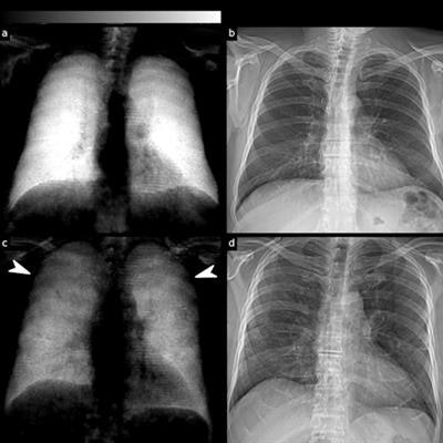
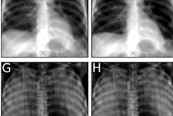
 (a) Dark-field and (b) conventional (attenuation-based) chest radiographs of a healthy subject. The dark-field radiograph exhibits a strong, homogeneous dark-field signal. The respective attenuation-based image shows no apparent pathology. (c) Dark-field and (d) attenuation-based chest radiographs of a patient infected with COVID-19. Compared with the healthy subject, the infected patient shows an overall decrease of signal intensity. While the signal of the healthy subject is homogeneous, the dark-field signal of the infected patient appears inhomogeneous and patchy, especially in the periphery of the lung (arrowheads). Image courtesy of Research Square.
In a receiver operating characteristic (ROC) analysis for the differentiation between infected patients and healthy subjects, the effect size expressed as area under the ROC curve (AUC) was 0.78 for standard radiographs, 0.91 for dark-field images, and 0.93 for the combination of both.
"In this study we present the first application of the recently developed dark-field x-ray imaging technology for the assessment of COVID-19-pneumonia and demonstrate its superiority over conventional radiography," the authors wrote.
Ultimately, the researchers are aiming to introduce a low-radiation, medical imaging alternative to CT imaging for COVID-19 pneumonia detection and follow-up. Although the sensitivity radiologists achieved when reading both dark-field chest x-rays and conventional chest x-rays together in this study was not as high as seen in CT imaging, it is still reasonably high and comes with only a fraction of the CT radiation dose, according to the authors.
However, further technological improvements are required, as well as clinical studies to evaluate the prototype's potential for lung imaging, the researchers wrote.
"Dark-field imaging might be a low-radiation alternative for disease monitoring especially in patients where repetitive CT scans should be avoided," Frank and colleagues concluded.
![Examples of ultrasound findings and techniques. (A) Images in a 39-year-old male patient with a mass in the left thigh. The mass is heterogeneous on the B-mode US image (compared with the patient in D) and showed increased microvascularity (superb microvascular imaging [SMI]) and shear-wave elastography (SWE) values. Undifferentiated pleomorphic sarcoma was diagnosed at biopsy (with pleomorphic rhabdomyosarcoma in surgical specimen). (B) Images in an 18-year-old male patient with a mass in the left leg. The mass is hypoechoic on the B-mode image, with no other findings suggestive of malignancy. The lesion is in contact with the cortex of the tibia, which is slightly irregular. CT revealed a doubtful anteromedial tibial erosion. The microvascular study demonstrated high vascularization, suggestive of malignancy. Periosteal Ewing sarcoma was diagnosed with both histologic and immunohistochemical confirmation. (C) Images in a 69-year-old female patient with a lump growing on the outside of the left leg. Multiple SWE examinations were performed (please note the high values obtained in the measurements, whereas the color map highlights the stiffness relative to adjacent tissues). SMI showed areas of increased vascularization to target for sampling. Undifferentiated spindle cell sarcoma was diagnosed at biopsy, with residual leiomyosarcoma in the surgical specimen after neoadjuvant therapy. (D) Images in a 56-year-old female patient with a mass in the right thigh. The mass is heterogeneous at both B-mode ultrasound (similar to patient A) and MRI (coronal T2-weighted spectral attenuated inversion recovery [SPAIR]; T1-weighted pre-contrast and postcontrast imaging), which even shows uptake after the administration of paramagnetic contrast material, which is traditionally suggestive of malignancy. Low values at SMI and elastography are suggestive of benignity. Spindle cell lipoma was diagnosed at biopsy, with atypical spindle cell lipomatous tumor in the surgical specimen.](https://img.auntminnie.com/mindful/smg/workspaces/default/uploads/2026/08/images-radiol250278fig2.APCFLSvX6p.jpg?auto=format%2Ccompress&fit=crop&h=100&q=70&w=100)



![Examples of ultrasound findings and techniques. (A) Images in a 39-year-old male patient with a mass in the left thigh. The mass is heterogeneous on the B-mode US image (compared with the patient in D) and showed increased microvascularity (superb microvascular imaging [SMI]) and shear-wave elastography (SWE) values. Undifferentiated pleomorphic sarcoma was diagnosed at biopsy (with pleomorphic rhabdomyosarcoma in surgical specimen). (B) Images in an 18-year-old male patient with a mass in the left leg. The mass is hypoechoic on the B-mode image, with no other findings suggestive of malignancy. The lesion is in contact with the cortex of the tibia, which is slightly irregular. CT revealed a doubtful anteromedial tibial erosion. The microvascular study demonstrated high vascularization, suggestive of malignancy. Periosteal Ewing sarcoma was diagnosed with both histologic and immunohistochemical confirmation. (C) Images in a 69-year-old female patient with a lump growing on the outside of the left leg. Multiple SWE examinations were performed (please note the high values obtained in the measurements, whereas the color map highlights the stiffness relative to adjacent tissues). SMI showed areas of increased vascularization to target for sampling. Undifferentiated spindle cell sarcoma was diagnosed at biopsy, with residual leiomyosarcoma in the surgical specimen after neoadjuvant therapy. (D) Images in a 56-year-old female patient with a mass in the right thigh. The mass is heterogeneous at both B-mode ultrasound (similar to patient A) and MRI (coronal T2-weighted spectral attenuated inversion recovery [SPAIR]; T1-weighted pre-contrast and postcontrast imaging), which even shows uptake after the administration of paramagnetic contrast material, which is traditionally suggestive of malignancy. Low values at SMI and elastography are suggestive of benignity. Spindle cell lipoma was diagnosed at biopsy, with atypical spindle cell lipomatous tumor in the surgical specimen.](https://img.auntminnie.com/mindful/smg/workspaces/default/uploads/2026/08/images-radiol250278fig2.APCFLSvX6p.jpg?auto=format%2Ccompress&dpr=2&fit=crop&h=167&q=70&w=250)


