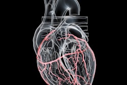
A software-based method that calculates fractional flow reserve from coronary angiography images has been validated in patients with intermediate coronary artery lesions, according to a study published March 29 in the New England Journal of Medicine.
In a clinical trial (FAST III), vessel fractional flow reserve (vFFR) was a noninferior alternative to invasive pressure-wire–based FFR for guiding revascularization decisions across more than 2,211 patients at 37 European centers, noted lead author Joost Daemen, MD, of Erasmus University Medical Center in Rotterdam, the Netherlands, and colleagues.
"The use of vFFR eliminates the need for guiding catheters, invasive coronary artery instrumentation and hyperemic agents with inherent risks and patient discomfort," Daemen said in a press release.
Guidelines for the management of coronary artery disease recommend the use of fractional flow reserve (FFR) to guide revascularization of intermediate coronary-artery lesions, the authors explained. Pressure-wire–based FFR is the standard technique, yet involves threading a pressure wire into the coronary artery and administering adenosine to induce hyperemia, which adds time, cost, and patient discomfort, they noted.
Alternatively, angiography-based vFFR (Caas vFFR, Pie Medical Imaging, the Netherlands) derives an FFR equivalent from two standard angiographic projections using computational fluid dynamics. In previous studies, the less invasive technique has shown diagnostic accuracy similar to pressure-wire–based FFR, and in this trial the researchers assessed whether it is noninferior to FFR-guided revascularization with respect to death from any cause, myocardial infarction, or revascularization.
Between November 2021 and May 2024, the researchers enrolled 1,116 patients in the vFFR group and 1,095 in the FFR group. All patients had chronic or acute coronary syndromes and at least one intermediate coronary lesion.
At one year, a primary end-point event had occurred in 80 patients (Kaplan–Meier estimate, 7.5%) in the vFFR group and in 79 patients (Kaplan–Meier estimate, 7.5%) in the FFR group (risk difference, −0.02 percentage points; 95% confidence interval, −2.25 to 2.21; p = 0.004 for noninferiority). vFFR flagged a higher proportion of lesions as hemodynamically significant -- 40.9% versus 31.3% with FFR -- which led to more patients undergoing revascularization in the vFFR group (45% versus 36%), the researchers reported.
In addition, the mean procedure time was modestly shorter in the vFFR group at 55.8 minutes compared to 60.9 minutes, and intraprocedural complications at the time of physiological assessment were also lower with vFFR than FFR (3.7% versus 6%), which the authors attributed to the absence of coronary wire manipulation in the vFFR arm.
“[vFFR] may streamline procedures and allows the use of physiology in centers in which only diagnostic coronary angiography is performed and may help to guide heart team decision making,” Daemen said.
The trial was sponsored by the European Cardiovascular Research Institute and co-funded by Pie Medical Imaging and Siemens Healthineers, which markets vFFR as part of its angiography platform.
The investigators called for longer follow-up to assess durability of outcomes and noted that they plan to conduct a cost-effectiveness analysis.
"The results of this trial contribute to the knowledge base of physiology-guided revascularization," the authors concluded.
The full study is available here.