PET imaging shows promise as a tool for monitoring disease activity in patients with autoimmune encephalitis, according to a recent study.
In 25 participants, researchers in Munich used a novel PET radiotracer called F-18 fluorodeprenyl-D2 (F-18 F-DED) to image reactive astrogliosis, a feature commonly observed in the disease, noted lead author Julia Dorneich of LMU University Hospital and colleagues.
“F-18 F-DED PET represents an encouraging imaging modality for in vivo assessment and monitoring of astrogliosis,” the group wrote. The study was published March 24 in Annals of Clinical and Translational Neurology.
Autoimmune encephalitis is a serious condition where the immune system attacks the brain, causing inflammation, cognitive decline, and psychiatric symptoms. Overactivated astrocyte cells (astrogliosis) are key players in the process, the researchers explained.
Recently, the group demonstrated the utility of F-18 F-DED PET for monitoring regional astrogliosis in patients with the second most common subtype of the disease called LGI1-antibody encephalitis. In this study, they aimed to further assess the technique in patients with the most common subtype, namely anti-glutamic acid decarboxylase 65 (anti-GAD65) encephalitis.
The investigators enrolled 25 patients -- 17 with limbic encephalitis/temporal lobe epilepsy (LE/TLE), four with cerebellar ataxia, and four with stiff-person syndrome, with overlapping phenotypes in nine -- as well as eight controls. PET scans were acquired (Biograph 64, Biograph mCT, Siemens Healthineers) over 60 minutes. The researchers assessed global astrogliosis by volumes of distribution (VT) and correlated PET measures with clinical phenotypes.
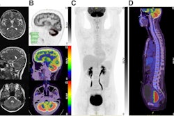
 (A) Overview of study participants with different clinical phenotypes depicted in distinct colors: Limbic encephalitis (LE)/temporal lobe epilepsy (TLE): Blue; cerebellar ataxia (CA): Green; stiff person syndrome (SPS): Red, including overlapping clinical features with an overview of the analyzed parameter; generated in Biorender. (B) F-18 F-DED uptake, as visualized by SUVr with global mean scaling, is shown for three representative patients with GAD65-AIE: Patient #6 with LE/TLE, showing mesiotemporal F-18 F-DED uptake; patient #16 with overlapping clinical features of CA, SPS, and LE, showing both mesiotemporal and cerebellar F-18 F-DED uptake; and patient #17 with isolated SPS showing no increased F-18 F-DED uptake. Additionally, the average F-18 F-DED uptake of eight controls is shown on the right.Annals of Clinical and Translational Neurology
“F-18 F-DED PET reveals region-specific astrogliosis corresponding to clinical manifestations and disease severity in GAD65-[encephalitis],” the investigators wrote.
They noted that the onset of autoimmune encephalitis may be subacute or insidious and that treatment responses to immunotherapy are often incomplete. Thus, many patients develop a chronic disease course with persistent deficits and fluctuating activity, they wrote. Furthermore, currently available paraclinical markers are limited.
Beyond aiding clinical assessment and phenotype classification, F-18 F-DED PET could be a tool for monitoring disease activity and treatment response, as it reflects disease severity in LE/TLE and cerebellar ataxia, the researchers suggested.
“However, prospective studies will be necessary to adequately assess and evaluate the applicability for monitoring the response to immunotherapy,” the group concluded.
The full study is available here.