A new study published in Scientific Reports demonstrates that F-18 FAPI-PET/CT detects significantly more hepatocellular carcinoma lesions than traditional F-18 FDG-PET/CT across all disease sites, offering superior imaging for HCC staging and treatment planning.
- F-18 FAPI-PET/CT achieved 96.7% detection rate for primary intrahepatic lesions compared to 60.0% with FDG-PET/CT
- FAPI-PET showed higher detection rates for lymph node metastases (94.6% vs 80.4%) and distant metastases (78.8% vs 48.5%)
- FAPI tracers target fibroblast activation protein (FAP), bypassing FDG-PET limitations like poor uptake and high liver background signal
- FAPI-informed management changes were implemented in 57.9% of patients, with staging changes in 19 of 32 initial staging cases
- Study enrolled 38 pathologically confirmed HCC patients in a prospective trial from May 2023 to June 2025
F-18 fibroblast activation protein inhibitor (FAPI) PET/CT detected more hepatocellular carcinoma (HCC) lesions than F-18 FDG-PET/CT at every disease site, according to a study published October 20 in Scientific Reports.
A group from China-Japan Union Hospital of Jilin University in Changchun, China, performed a prospective head-to-head comparison of the two techniques in patients with pathologically confirmed disease, with findings suggesting FAPI-PET offers additional clinical value for staging and treatment-planning.
"F-18 FAPI-PET/CT showed higher lesion detection in selected HCC settings than F-18 FDG-PET/CT for primary intrahepatic, lymph-node, and distant metastatic lesions and may provide complementary information for staging and treatment planning," the group wrote.
There is a pressing need for HCC imaging agents with higher specificity and sensitivity, as FDG-PET/CT’s utility in HCC is hampered by poor FDG uptake and rapid tracer clearance, compounded by the liver’s own high background FDG signal, the group wrote. FAPI tracers bypass those limitations by targeting fibroblast activation protein (FAP), which is highly expressed in the cellular microenvironment surrounding HCC tumors, they added. Prior studies suggest FAPI-PET/CT may be superior to FDG-PET/CT, and in this study the researchers sought to build on the evidence.
The researchers enrolled 38 patients with pathologically confirmed HCC in a prospective trial from May 2023 to June 2025. All underwent paired F-18 FDG-PET/CT and F-18 AlF-NOTA-FAPI-04 PET/CT within seven days.
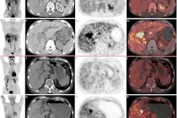
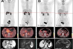
 A 71-year-old female patient presented with abdominal pain and distension for over two months. Non-contrast CT revealed multiple hypodense lesions in the liver. (a) F-18 FAPI- PET/CT demonstrated intensely elevated tracer uptake in the hypodense hepatic lesions (arrowhead; SUVmax = 8.92), which were pathologically confirmed as HCC with multiple
intrahepatic metastases. Additionally, metastases were detected in the hepatic hilar and cardiophrenic angle LNs (dotted
arrowheads; SUVmax = 8.02). (b) F-18 FDG PET/CT showed lower uptake in the intrahepatic lesions compared to F-18 FAPI
(arrowhead; SUVmax = 4.95). Similarly, the LN metastases (dotted arrowheads; SUVmax = 3.58) also exhibited reduced uptake.Scientific Reports
Additionally, relative to F-18 FDG, F-18 FAPI-PET/CT produced staging changes in 19 of 32 patients undergoing initial staging, including six true upstagings, and a FAPI-informed management change was implemented in 57.9% (22/38) of patients, the group reported.
Although the post-FAPI treatment strategies were implemented, the present study was not designed to determine whether FAPI-informed management changes improved patient outcomes, the researchers noted.
"Prospective longitudinal studies with extended follow-up periods are warranted to establish the prognostic significance of F-18 FAPI-based quantitative parameters in predicting therapeutic outcomes and long-term survival," the authors wrote.
The full study is available here.