A higher prevalence of intracranial aneurysms is associated with greater cumulative combat blast exposure among special operations military forces, according to a study published April 14 in Radiology.
A team led by Sara De Giorgi, MD, of Massachusetts General Hospital in Boston, found that intracranial aneurysms were three times more common in highly exposed personnel.
"Even after accounting for other health factors such as age and blood pressure, the association remained significant," she said in a statement released by the RSNA. "These findings suggest that repeated blast exposure may leave a measurable vascular signature in the brain."
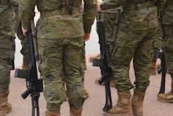
Special operations forces personnel are trained for clandestine, high-risk, and time-sensitive missions, and specialize in unconventional warfare, direct action, counterterrorism, and reconnaissance. They include the Green Berets, 75th Ranger Regiment, Navy SEALs, and the Marine Forces Special Operations Command, and are directed by U.S. Special Operations Command.
These personnel are often exposed to blast forces, but the long-term effects of these injuries on the brain aren't clear, the group explained. To address this knowledge gap, the researchers conducted a study that included 564 special operations forces personnel who underwent 3-tesla brain MRI with time-of-flight MR angiography (MRA); the group then assessed the prevalence of structural brain MRI abnormalities and examined any association with cumulative blast exposure. The researchers quantified blast exposure using the Generalized Blast Exposure Value (GBEV) -- a numerical score between one and 10 that measures the cumulative impact of repeated low-intensity blasts (e.g., from training; GBEV high exposure, ≥6.55, and low exposure, <6.55). Mean age of the study participants was 43 years.
De Giorgi and colleagues reported the following:
- The most prevalent MRI finding was white matter hyperintensities (215/564, 38.1%), followed by intracranial aneurysms (33/564, 5.9%).
- Among all evaluated MRI findings, only intracranial aneurysms were associated with cumulative blast exposure (mean GBEV in participants with aneurysms, 7.02, compared to 6.54 in those without aneurysms; p = 0.01).
- Aneurysm prevalence was greater in the high-exposure group (21/220, 9.5%) compared to the low-exposure group (6/220, 2.7%).
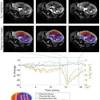
 Representative axial time-of-flight MR angiography image in a 39-year-old male participant shows a laterally projecting intracranial aneurysm (arrow) originating from the right cavernous segment of the internal
carotid artery.RSNA
The findings suggest a possible long-term vascular effect of repeated low-level blast exposure during years of service, De Giorgi noted.
"These vascular changes can be seen with routine MRI scans, making the findings directly relevant to everyday radiology practice," she said. "Radiologists may use this information when interpreting brain MRIs in patients with a history of repeated blast exposure, helping identify possible vascular abnormalities, such as aneurysms. In addition, our preliminary results suggest that screening MRAs may be warranted in this population."
The study is "necessary and timely," wrote Pejman Jabehdar Maralani, MD, and colleague Vivek Pai, MD, both of the University of Toronto in Canada, in an accompanying editorial.
"De Giorgi et al provide a valuable addition in the ongoing efforts to create normative data benchmarks for military personnel, which can ultimately be applied to nonmilitary individuals," they concluded.
Access the full study here.