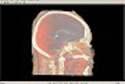
A short or absent nasal bone as noted on second-trimester ultrasound may indicate a fetus with Down syndrome, according to a new study.
Dr. Beryl Benacerraf, clinical professor of radiology and obstetrics and gynecology at Harvard Medical School, presented initial findings from her group’s research at the 2002 Society of Radiologists in Ultrasound meeting in San Francisco. The study will be published in the December issue of the Journal of Ultrasound in Medicine; the first co-author is Dr. Bryann Bromley, an associate clinical professor of obstetrics, gynecology and reproductive biology, also at Harvard Medical School in Boston.
A number of obstetrical ultrasound findings have been previously identified in the medical literature as frequent but nondefinitive markers of Down syndrome in the second trimester. These include nuchal translucency indicating redundant skin at the back of the neck, hyperechoic bowel, short femur and humerus bones, and others.
Researchers have only recently focused on nasal bone length as a possible sonographic marker, even though Dr. J. Langdon Down in 1866 described a characteristically small nose as part of the syndrome. In 2001, British researchers looking at first-trimester sonograms reported that a short or missing nasal bone identified 73% of Down syndrome fetuses with a 1% false-positive rate (The Lancet, November 17, 2001, Vol.358:9294, pp. 1665-1667).
More recently, researchers from Argentina found that the absence of nasal bone on first-trimester ultrasound was significantly associated with Down syndrome. Their study was published in Prenatal Diagnosis (October 2002, Vol. 22:10, pp.930-932).
"Having seen that we wanted to evaluate nasal bone length as a sonographic marker in the second trimester," Benacerraf said, "because, at least in Boston, we see most of our patients coming in for their obstetrical ultrasound mostly in the second trimester."
"If you get a very good sagittal image, you will be able to identify the ossification of the nasal bone quite nicely as a finite measurement you can make," Benacerraf said. The researchers used electronic calipers to measure from the base of the nasal bone closest to the frontal bone to the distal extent of the ossification.
The prospective study evaluated 239 consecutive fetuses between 15 and 20 weeks gestation. The fetuses were already scheduled for an ultrasound and subsequent amniocentesis because of high-risk factors such as advanced maternal age and serum biochemical screening results.
Of the 239 fetuses examined, 16 were confirmed at amniocentesis as having Down syndrome. The 16 fetuses had a mean gestational age of 17 weeks and mothers a mean age of 37 years -- nearly identical demographics to those of the 223 normal euploid fetuses.
However, there was a statistically significant difference between the groups in their mean nasal bone length as measured at the second-trimester ultrasound. Among the fetuses with Down syndrome, the mean NBL was 3.5 mm, compared to 4.6 mm for those without Down syndrome, Benacerraf said.
To account for growth of the nasal bone with advancing gestational age, the researchers also calculated the ratio of NBL to biparietal diameter. That ratio ended up as a mean of 11.3 for the Down syndrome fetuses versus 8.1 for the euploid fetuses.
"This ratio among normal fetuses is pretty much constant throughout the gestational age window that we use, which is actually very helpful," Benacerraf said. "It therefore suggests that a single cutoff can be used to define abnormal regardless of this gestational age."
Using an NBL/BPD ratio of 10, the researchers were able to identify 81% of the fetuses with Down syndrome with an 11% false-positive rate. Using a lower ratio of 9 would have identified all of the Down syndrome fetuses but generated too many false positives, Benacerraf said.
Interestingly, 6 of the 16 Down syndrome fetuses, or 37%, had no visible nasal bone ossification at all, while that was the case for only one, or 0.5%, of the 223 euploid fetuses. Based on these findings, an absent nasal bone would therefore have a likelihood ratio of 83 -- making it a more accurate reported predictor of Down syndrome than any other sonographic marker in the second trimester, Benacerraf said.
A majority of the Down syndrome fetuses -- 13 of the 16 -- also exhibited other sonographic markers of aneuploidy. Of the three fetuses with no additional markers, two had absent nasal bones, she said.
"The presence of a nasal bone is an important new marker in the detection of fetuses with Down syndrome, which has the potential of being as accurate or as helpful as the nuchal fold," Benacerraf concluded.
The marker could help high-risk women avoid amniocentesis or identify possible aneuploid fetuses in low-risk women. However, Benacerraf added, "the significance of the nasal bone as an isolated finding, particularly in a low-risk population, has not yet been established."
By Tracie L. ThompsonAuntMinnie.com contributing writer
November 19, 2002
Related Reading
Fetal nasal bone appearance on ultrasound may guide Down syndrome detection, November 8, 2002
Turf Wars in Radiology IV: Radiologists, ob/gyns sound off on fetal imaging, September 26, 2002
Newer Down's syndrome screening tests do not improve detection rate, July 9, 2002
Genetic ultrasound capable of preventing amniocentesis losses, July 8, 2002
For perinatal diagnosis of Down syndrome, ultrasound ups the odds, January 4, 2002
Copyright © 2002 AuntMinnie.com


