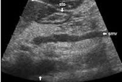
Examining the pancreas with ultrasound can be a challenging prospect, due to the complexity of the organ and surrounding tissues. But with training, education, and experience, ultrasound professionals can learn to use anatomic landmarks to improve their visualization of the organ.
That’s the message from Dr. Lars Thorelius of University Hospital in Linköping, Sweden. In the third installment of his ongoing series of articles for AuntMinnie.com, Dr. Thorelius has produced a guide to the proper examination techniques for imaging the normal pancreas.
As in Dr. Thorelius’ previous contributions, this week’s article employs an image-compression technique that enables dynamic ultrasound clips to be viewed over the Internet using off-the-shelf media players. We’re featuring the piece in our Ultrasound Digital Community, at http://ultrasound.auntminnie.com.
In addition to the dynamic clips and still images, the article includes a special section on common mistakes experienced when evaluating the pancreas. For example, students trying to access hidden parts of the pancreas shouldn’t hesitate to get patients out of the supine position when they can’t find what they’re looking for. To learn about other pancreatic pitfalls, just click here.


