Sunday, November 25 | 11:15 a.m.-11:25 a.m. | SSA20-04 | Room S102CD
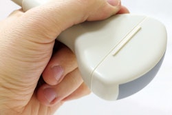
Ultrasound tomography is better than handheld ultrasound when it comes to measuring breast tumor volume, researchers will report in this presentation.Accurately measuring lesion volume helps clinicians better diagnose and treat breast cancer, wrote a team led by Dr. Rajni Natesan of MD Anderson Cancer Center in Houston.
"Since tumor volume doubling time is associated with growth rate and tumor biology, accurate measurement of tumor volume is critical for oncologic diagnosis, staging, and treatment," the researchers wrote.
Ultrasound tomography produces 3D sound maps that identify tissue types and measure lesion volumes. For the study, the group used six cylindrical phantoms embedded with "lesions" with known volumes ranging from 1.3 cm3 to 7.4 cm3. The researchers imaged the phantoms with both ultrasound tomography and handheld ultrasound; two radiologists then interpreted the images.
Natesan's group found that ultrasound tomography's volume estimates were congruent with the actual volumes of the lesions, while handheld ultrasound volumes were significantly larger than the true volumes, with a mean overestimation of 62.8% for spherical phantoms and 32.6% for ellipsoid phantoms.
"Ultrasound tomography can accurately measure the volume of irregular-shaped masses, with superior accuracy than handheld ultrasound," the group concluded.


