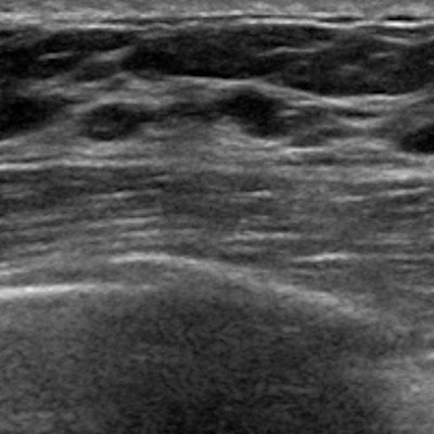
Contrast-enhanced ultrasound (CEUS) can distinguish ductal carcinoma in situ (DCIS) from fibroadenoma in breast tissue, according to a study published online June 17 in the Journal of Ultrasound in Medicine.
The findings further support the use of CEUS in the breast cancer screening arsenal, especially since DCIS can be tough to diagnose, wrote a team led by Weiwei Li of Shanghai Jiao Tong University.
"Ultrasound has become an important screening tool for evaluating breast lesions because of its widespread availability, low cost, and safety," the group wrote. "CEUS ... has been shown to be very helpful for differentiating benign from malignant breast tumors."
DCIS is a precursor to invasive breast cancer, Li's group noted. But accurately diagnosing DCIS -- and distinguishing it from benign lesions like fibroadenomas -- can be tricky. Quantitative breast ultrasound has been used to do this, but the modality has its limitations.
That's where CEUS comes in, since it can evaluate blood distribution and provide qualitative and quantitative analyses for characterizing breast lesions, the researchers wrote.
"Contrast-enhanced ultrasound is a noninvasive method that uses microbubble agents to evaluate and quantify the tissue perfusion," the group wrote. "Hence, it is reasonable to assume that the different enhancement characteristics may be used to evaluate the microcirculation perfusion, as well as to offer more valuable information for distinguishing malignant breast lesions from benign ones."
However, no studies to date have explored the diagnostic value of contrast ultrasound for identifying DCIS, according to the authors. So Li and colleagues assessed the characteristics of DCIS on CEUS, comparing CEUS findings of DCIS and fibroadenoma.
Their study included 251 women with 127 histopathologically confirmed DCIS lesions (mean size, 26 mm) and 124 fibroadenoma lesions (mean size, 29 mm). All of the women had undergone CEUS. Li and colleagues then analyzed the CEUS findings of the DCIS and fibroadenomas for distinguishing features.
The group found that DCIS lesions more often had particular features than the fibroadenomas and that CEUS could identify them. These included the following:
| Features identified by CEUS as characteristic of DCIS | ||
| Feature | DCIS | Fibroadenomas |
| Blood perfusion defects | 74.6% | 42.6% |
| Earlier wash-in time | 65.9% | 38.5% |
| Earlier wash-out time | 38.9% | 26.2% |
| Hyperintense enhancement | 44.4% | 20.5% |
| Penetrating vessels | 70.6% | 41.8% |
The results confirm that CEUS offers clinicians an effective way to diagnose DCIS, Li and colleagues concluded.
"As a novel diagnostic modality, CEUS can be used to evaluate the microvessels of DCIS with a low volume and blood flow velocity," the group wrote. "The features, including an earlier wash-in time, hyperintense enhancement, blood perfusion defects, an enlarged enhancement scope, and penetrating vessels, are more frequent in DCIS than in fibroadenoma. Thus, CEUS may be used as a valuable tool for differential diagnosis of DCIS and fibroadenoma lesions."


