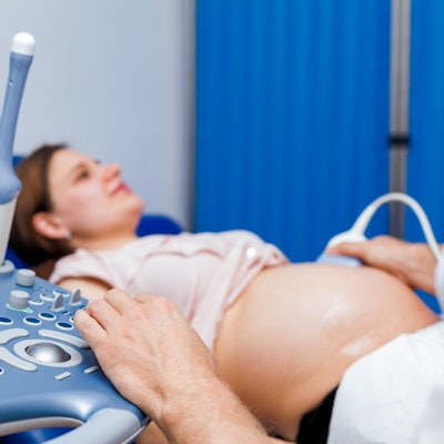
The ultrasound probes and systems that ob/gyn professionals typically rely on for prenatal checkups can also be used for lung imaging in patients with suspected cases of COVID-19, according to an article published on March 24 in Ultrasound in Obstetrics & Gynecology.
The use of lung ultrasound scans can help identify pregnant patients with early stages of COVID-19 as well as those who may have had a false-negative result from a polymerase chain reaction (PCR) test. In addition, ob/gyn clinicians already have ultrasound experience and equipment in their practices.
"Lung ultrasound can be considered as an 'extension' of the obstetric abdominal ultrasound evaluation," wrote the authors, led by Dr. Francesca Moro, PhD, from Fondazione Policlinico Universitatrio A. Gemelli in Rome. "The ultrasound examiner can simply move the probe from the abdomen to the chest, scanning the anterior and lateral areas of the thorax."
While CT has excellent ability to detect COVID-19, concerns remain regarding radiation exposure for pregnant women. The authors proposed using lung ultrasound scans for these women since ob/gyns already use ultrasound and the modality has demonstrated its usefulness in diagnosing early-stage COVID-19 pneumonia.
To perform lung ultrasound scans on pregnant patients, the authors first recommend scanning the basal to upper zones of the thorax. This can be done by following four vertical lines: right midaxillary line, right parasternal line, left midaxillary line, and left parasternal line. Then, the clinician should ask the patient to move to a lateral or sitting position in order to scan their back in a similar pattern.
"With the patient then in a sitting or lateral position, the posterior paravertebral surface of the thorax should be scanned, from the basal to upper zones or posterior-axillary lines according to the patient's position," they wrote.
In a normal lung ultrasound scan, the only visible findings are the pleural line and A-lines, which are caused by the reflections of the ultrasound beam. But in a lung scan of a patient with COVID-19, B-lines are also present, as well as subpleural consolidations, a thickened or patchy pleural line, and other structures that can indicate pneumonia and advanced respiratory distress syndrome.
Any type of ultrasound machine, including older models, can be used to perform lung ultrasound scans, the authors noted. However, handheld ultrasound scanners may be best suited to sterilizing and covering the instruments in an emergency.
In addition, no ultrasound probes are specifically designed for lung imaging. The authors recommended using a linear probe for evaluating the pleural line, which is consistent with recommendations from other researchers. They also noted convex probes, which are already common in ob/gyn practices, can be used for lung imaging as well.
Finally, the authors proposed several focus and gain settings. They advised adjusting the focus to a level consistent with a sufficient view of the pleural line and recommended reducing the gain to highlight A-lines, B-lines, and other hyperechoic signs. However, if a patient has a consolidated lung, the gain may have to be adjusted for that used for the organ parenchyma.
"We have proposed a systematic approach for obstetricians/gynecologists to perform lung ultrasound examination in pregnant women, describing potential applications and semiology and providing practical points for consideration," the authors concluded. "Pathological ultrasound patterns are compared to those expected in a normal lung, with particular emphasis on those more indicative of COVID-19 infection."


