Clarius Mobile Health is highlighting new features for its ultrasound product suite, including enterprise software for the company's scanners, new capabilities powered by AI, and built-in CPT medical codes to help with billing and reimbursement.
The company said these features aim to improve workflow, streamline billing, and make handheld ultrasound easier to learn and use. The features, some of which can be accessed via the Clarius App 12.0, will be available on the Apple and Google Play stores to Clarius users starting January 21.
The features include the following:
- AI-powered T-Mode can provide real-time guidance with AI-powered colored and textured anatomical overlays to improve learning of ultrasound grey scale images, now available for imaging the rotator cuff.
- Through the app, clinicians billing for musculoskeletal (MSK), urology, and women's health exams can select the right CPT code to streamline workflow for billing and reimbursements.
- A-Mode allows for ophthalmologists to use Clarius ultrasound to measure the axial length of the eye to help with diagnosis and monitoring of conditions such as myopia, hyperopia, and cataracts. It is also valuable for assessing retinal and choroidal pathologies.
- A new protocol introduced by Clarius allows clinicians to change settings to optimize facial scans and reduce interruptions during patient consultations for aesthetic procedures.
- Through the app, clinicians can use a 12-field lung assessment with scoring from 0 to 3 and a cumulative total provided to support clinical decision-making at the point of care.
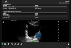
The company added that Clarius users in Australia, Iran, and other select regions can now access new AI-powered features designed to increase the efficiency of clinicians while performing ultrasound exams with Clarius handheld ultrasound scanners. These include MSK AI for key anatomical structures, OB AI to streamline fetal measurements, and Bladder AI to provide bladder volume data.







![Examples of ultrasound findings and techniques. (A) Images in a 39-year-old male patient with a mass in the left thigh. The mass is heterogeneous on the B-mode US image (compared with the patient in D) and showed increased microvascularity (superb microvascular imaging [SMI]) and shear-wave elastography (SWE) values. Undifferentiated pleomorphic sarcoma was diagnosed at biopsy (with pleomorphic rhabdomyosarcoma in surgical specimen). (B) Images in an 18-year-old male patient with a mass in the left leg. The mass is hypoechoic on the B-mode image, with no other findings suggestive of malignancy. The lesion is in contact with the cortex of the tibia, which is slightly irregular. CT revealed a doubtful anteromedial tibial erosion. The microvascular study demonstrated high vascularization, suggestive of malignancy. Periosteal Ewing sarcoma was diagnosed with both histologic and immunohistochemical confirmation. (C) Images in a 69-year-old female patient with a lump growing on the outside of the left leg. Multiple SWE examinations were performed (please note the high values obtained in the measurements, whereas the color map highlights the stiffness relative to adjacent tissues). SMI showed areas of increased vascularization to target for sampling. Undifferentiated spindle cell sarcoma was diagnosed at biopsy, with residual leiomyosarcoma in the surgical specimen after neoadjuvant therapy. (D) Images in a 56-year-old female patient with a mass in the right thigh. The mass is heterogeneous at both B-mode ultrasound (similar to patient A) and MRI (coronal T2-weighted spectral attenuated inversion recovery [SPAIR]; T1-weighted pre-contrast and postcontrast imaging), which even shows uptake after the administration of paramagnetic contrast material, which is traditionally suggestive of malignancy. Low values at SMI and elastography are suggestive of benignity. Spindle cell lipoma was diagnosed at biopsy, with atypical spindle cell lipomatous tumor in the surgical specimen.](https://img.auntminnie.com/mindful/smg/workspaces/default/uploads/2026/08/images-radiol250278fig2.APCFLSvX6p.jpg?auto=format%2Ccompress&fit=crop&h=112&q=70&w=112)


