Contrast-enhanced spectral mammography (CESM) can play a pivotal role in diagnosing some types of breast cancer, according to research presented at the RSNA 2020 virtual meeting. A new study found CESM yielded 93% accuracy for identifying high-grade ductal carcinoma in situ (DCIS).
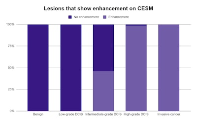
In the study of 510 cases, high-grade DCIS and invasive cancers almost exclusively showed enhancement on contrast-enhanced mammography. The findings reflect the pivotal role that CESM can play in further assessing suspicious microcalcifications on standard mammography, noted presenter Dr. Mohammed Mohamed Gomaa.
"The presence of associated underlying nonmass enhancement is an indicator of high-grade DCIS or invasiveness," said Gomaa, head of the radiology department at Baheya Charity Women's Cancer Hospital in Giza, Egypt, in an on-demand presentation.
All women in the study were between the ages of 27 and 77 and underwent standard mammography, which revealed suspicious microcalcifications. Gabaa and colleagues then used contrast-enhanced mammography to further visualize the suspicious microcalcifications.
The microcalcifications varied greatly in distribution and morphology on CESM. Grouped distribution and amorphous morphology were the most common presentations, but neither accounted for more than half of all cases.
Three-quarters of the 510 patients were eventually diagnosed with cancer. Of the 405 malignant lesions, 330 were DCIS and 75 were invasive cancers. The majority of DCIS cases (70%) were high grade, while 20% were low grade and 10% were intermediate grade.

Enhancement on CESM varied by the type of lesion. No benign or low-grade DCIS lesions showed enhancement on CESM, while all invasive cancers showed enhancement. Similarly, all but four high-grade DCIS lesions showed enhancement on CESM.
"Lack of enhancement is favorable to diagnose nonmalignant lesions or noninvasiveness or low-grade DCIS," Gomaa said.
CESM looked especially promising for identifying invasive or high-grade DCIS from suspicious microcalcifications. For high-grade DCIS, the modality yielded an accuracy of 93% and sensitivity of 98%. It also netted an accuracy of 88% and sensitivity of 99% for both invasive cancer and high-grade DCIS.
Despite its promising performance for higher-risk cancers, CESM only netted a positive predictive value of 62% and sensitivity of 79% for all lesion types combined. Still, Gomaa was excited by the modality's diagnostic performance potential.
He discussed several cases where CESM helped to distinguish between microcalcifications signaling cancer versus benign findings. In one case, a 44-year-old woman showed bilateral fibrocystic changes and rounded and punctate microcalcifications on mammography.
Although the microcalcifications appeared similar in each breast on standard mammography, further CESM revealed important differences. The patient's right breast showed nonmass enhancement on CESM, which corresponded to grade III invasive breast cancer. But the microcalcifications in the patient's left breast showed no enhancement on CESM and instead represented proliferative fibrocystic disease with usual duct hyperplasia.
"Contrast-enhanced spectral mammography has a pivotal role in assessment of grading and invasiveness of suspicious microcalcifications," Gabaa concluded.


