Minnies finalists, page 3
Scientific Paper of the Year
Accumulation of gadolinium in human cerebrospinal fluid after gadobutrol-enhanced MR imaging: A prospective observational cohort study. Nehra AK et al, Radiology, August 2018. To learn more about this paper, click here.
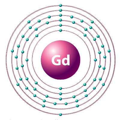
Given the major focus on gadolinium retention over the past year, it's no surprise that this paper made the final cut as one of two finalists for the Scientific Paper of the Year award in the Minnies.
Previous research has demonstrated that traces of gadolinium from MRI contrast can remain in the brain and other body tissues long after scans are performed. But many questions still remain, such as whether these gadolinium deposits are dangerous to patients, and whether different types of gadolinium-based contrast agents (GBCAs) are more prone to retention.
In the current study, Dr. Avinash Nehra and colleagues from the Mayo Clinic in Rochester, MN, used samples drawn from lumbar punctures to detect gadolinium in the cerebrospinal fluid of adult and pediatric patients after they received a macrocyclic GBCA. The trace elements were found even in patients who had normal renal function and an undamaged blood-brain barrier.
That's important, because previous speculation had theorized that gadolinium made its way into brain tissue only if the blood-brain barrier had been compromised. The findings by Nehra et al indicate that cerebrospinal fluid may be the route gadolinium follows to enter brain tissue -- even in healthy patients.
Interestingly, several co-authors on the current paper were also on the Mayo team that wrote the winning paper -- also on gadolinium retention -- in the 2015 Minnies awards. Will lightning strike twice for Mayo? Only the Minnies panel knows for sure ...
Thrombectomy for stroke at six to 16 hours with selection by perfusion imaging. Albers GW et al, New England Journal of Medicine, February 22, 2018. To learn more about this paper, click here.
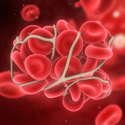
The emergency care of stroke patients has been revolutionized by the use of endovascular thrombectomy, an interventional procedure in which a stent-retrieval device is inserted under image guidance to capture and remove the clot and restore blood flow before brain tissue is further damaged. But endovascular thrombectomy is only recommended for patients within six hours of the onset of stroke.
As described in the current paper, researchers who were part of the Endovascular Therapy Following Imaging Evaluation for Ischemic Stroke 3 (DEFUSE 3) trial used software (Rapid, iSchemaView) to analyze blood perfusion on MRI and CT scans to identify stroke patients thought to have salvageable tissue. They found that performing thrombectomy up to 16 hours after stroke onset was effective, with more patients treated this way returning to functional independence, compared with a control group. These patients also had improved survival.
The results of DEFUSE 3 are significant because they extend the treatment window for many stroke patients, especially those who may have sustained a stroke in their sleep, which makes it difficult for emergency personnel to determine the time of stroke onset.
Albers assigns much of the credit for DEFUSE 3's findings to the Rapid software, which was initially developed at Stanford by Albers and co-researchers and licensed to iSchemaView. It was first used in the DEFUSE 2 trial, and it received U.S. Food and Drug Administration clearance in 2013.
Best New Radiology Device
Butterfly iQ personal ultrasound scanner, Butterfly Network
 Image courtesy of Butterfly Network.
This year's race for Best New Radiology Device has turned into an epic David versus Goliath contest in the ultrasound market segment, pitting start-up ultrasound developer Butterfly Network against medical imaging powerhouse GE Healthcare.
Butterfly was founded in 2011 by Jonathan Rothberg, PhD, a professor of genetics at Yale University who invented high-speed DNA sequencing. Rothberg wanted to develop technology to give more people around the world access to medical imaging, and he saw ultrasound as the potential solution due to its inherently low cost.
Five years later, the team Rothberg put together had developed what they call "ultrasound on a chip" -- a transducer that combines semiconductors, artificial intelligence, and cloud technology, all at the affordable price point of $1,999. The Butterfly iQ scanner combines functions that typically would require three ultrasound probes into a single ultrawide-band 2D matrix array that connects to an iPhone.
The Butterfly iQ transducer can be used for 13 clinical applications, and artificial intelligence can be employed to help physicians and less-skilled users interpret medical images. Butterfly iQ received FDA clearance in 2017, and in 2018 the company added a Tele-Guidance capability that enables remote experts to guide the use of the transducer in the field.
Rothberg believes that Butterfly iQ will revolutionize medical imaging in the same way that putting a camera on a semiconductor chip revolutionized photography. But it could also have major repercussions for medical imaging professionals by moving control over imaging away from trained healthcare professionals.
Logiq E10 ultrasound scanner, GE Healthcare
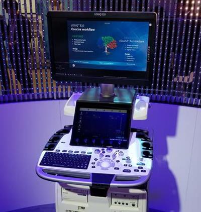 Logiq E10 ultrasound scanner.
And in this Minnies corner, we have the titan of radiology, GE Healthcare. GE is positioning Logiq E10 as its most advanced radiology ultrasound scanner ever, built from the ground up to support the emerging integration of artificial intelligence and medical imaging.
First introduced at the 2018 European Congress of Radiology (ECR), Logiq E10 is GE's flagship ultrasound scanner for radiology, shared services, and other clinical applications outside of the cardiology and ob/gyn segments. The system uses GE's cSound Architecture platform, which the company says acquires and reconstructs data in a manner similar to an MRI or CT system.
That requires a lot of data processing power, so GE built graphics processing units (GPUs) from NVIDIA into the Logiq E10 scanner. These GPUs will also enable E10 to support the growing use of artificial intelligence algorithms to analyze ultrasound images, the company believes.
Other features on Logiq E10 include a Photo Assistant App that allows users to photograph relevant anatomy and attach photos to clinical images that are sent to radiologists in the DICOM format. Another tool, Remote Clinical Application, enables users to manipulate scanner settings with a remote control on their tablet or smartphone. Cloud connections are made available via Tricefy from Trice Imaging.
Logiq E10 began shipping globally shortly after its ECR introduction.
Best New Radiology Software
Viz LVO CT artificial intelligence software, Viz.ai
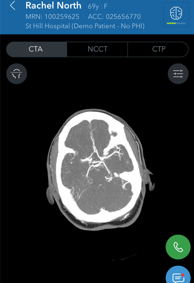 Image courtesy of Viz.ai.
Artificial intelligence (AI) software developer Viz.ai received U.S. Food and Drug Administration (FDA) marketing authorization in February for Viz LVO, an AI-based application that analyzes CT angiography images and notifies providers that their patient may be having a stroke.
After being notified of a suspected large vessel occlusion (LVO) stroke via a text message, neurologists and interventionalists can then view the patient's images, according to the vendor. Notably, Viz LVO was cleared early in 2018 by the FDA's via its "de novo" premarket review pathway, which allows vendors to petition the FDA to down-classify a novel device considered to have low to moderate risk and that had not previously been classified by the agency.
Traditionally, these applications would have been considered class III devices by default and would require the more rigorous premarket approval (PMA) process. The de novo action also established a new predicate device and regulatory classification, enabling subsequent computer-aided triage devices with the same intended medical imaging use to go through the 510(k) clearance process.
Horos Cloud Reporting, Horos Project
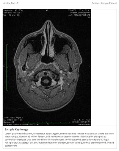 Image courtesy of Horos Project chief sponsor Purview.
The other finalist for Best New Radiology Software comes from the Horos Project, an open-source initiative to develop a 64-bit medical image viewer for Apple's OS X operating system. Horos Cloud Reporting, which was released by Horos chief sponsor and image management firm Purview in July, adds structured reporting capability to the popular open-source Horos viewer.
With Horos Cloud Reporting, physicians can create, save, and share customized structured reports as DICOM files within minutes, according to Purview. Horos Cloud Reporting is a secure platform that can be launched from within the Horos viewer, and it is available on a subscription basis for $1.99 per month. The Horos viewer is used by more than 125,000 professionals in over 170 countries, the company said.
Purview is currently pursuing regulatory approval for Horos in both the U.S. and elsewhere.
Best New Radiology Vendor
Ai Visualize

This artificial intelligence developer introduced itself to the medical imaging community at RSNA 2017 in Chicago, where it unveiled its cloud-based AI image analysis and viewing software.
The cloud visualization platform, which has been applied in other industries such as cloud 3D gaming, augmented reality, virtual reality, and autonomous driving, employs deep-learning algorithms to assess imaging data for diagnostically valuable information that is not apparent through conventional analysis, according to the vendor. It then also transmits detailed 3D/2D renderings to users on any internet-enabled device. Information and imaging tools are streamed in real-time to end users with the aid of predictive buffering.
The software supports x-ray, ultrasound, CT, MRI, 3D tomosynthesis, and digital pathology images. It has 11 international patents and received U.S. Food and Drug Administration clearance.
EnvoyAI
EnvoyAI, a spin-off of advanced visualization software developer TeraRecon, has the goal of creating a type of Amazon.com for artificial intelligence algorithms.
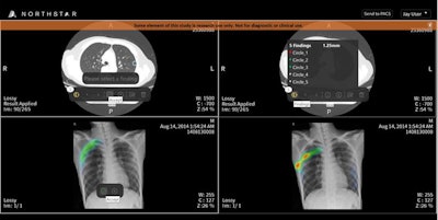 Several EnvoyAI platform results from AI companies on the EnvoyAI Exchange are shown within TeraRecon's Northstar AI Results Explorer. Image courtesy of TeraRecon.
Formerly known as McCoy Medical Technologies prior to its acquisition by TeraRecon in June 2017, EnvoyAI formally debuted at RSNA 2017 with EnvoyAI Exchange -- a vendor-neutral distribution platform designed to connect AI software developers with researchers and healthcare providers. Developers can use the Exchange to share their algorithms for testing and, later on, for clinical use and commercialization. Meanwhile, radiologists can use the EnvoyAI platform to interact with AI in their native workflow, according to the company.
As of August, the platform offered access to 71 AI applications from 30 development partners. Some of the algorithms have received U.S. Food and Drug Administration clearance and can be purchased by end users, while others are available for research use only.
Best Radiology Mobile App
CTisus iQuiz: The HD Edition, Dr. Elliot Fishman (iOS)
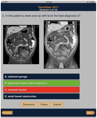 CTisus iQuiz: The HD Edition. Image courtesy of Dr. Elliot Fishman.
Dr. Elliot Fishman of Johns Hopkins University is in contention for what would be his third straight Minnie in the Best Radiology Mobile App category. He won in 2017 for CTisus iPearls and in 2016 for CTisus Critical Diagnostic Measurements. Another member of the CTisus family of apps -- CTisus iLecture Series -- was a finalist in 2014.
This year's finalist, iQuiz: The HD Edition, features more than 1000 cases in 15 different categories that are presented in a quiz format. Each case includes specific "pearls" -- or bits of knowledge -- as well as an audio discussion. Version 9, released earlier this year, added six new quizzes for a total of 60 more cases.
Like all of the CTisus apps, iQuiz: The HD Edition is available for free.
Radiology Assistant 2.0, BestApps (iOS)
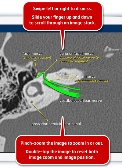 Radiology Assistant 2.0. Image courtesy of Dr. Woulter Veldhuis, PhD.
Fishman faces a familiar foe in the finals: Radiology Assistant 2.0 is a Best Radiology Mobile App finalist for the second year in a row. The initial version of the popular educational app debuted in 2010 and was also a finalist in the 2013 Minnies.
Launched in 2017, Radiology Assistant 2.0 continues to provide peer-reviewed articles from expert radiologists around the world -- but with a significantly improved user interface, according to Dr. Wouter Veldhuis, PhD, a radiologist at University Medical Center Utrecht in the Netherlands. The app was completely rewritten and rebuilt using Apple's Swift programming language, and it includes a new article layout and better searching capability. Users can view inline and full-screen movies and scroll through image stacks, for example. Offline access to articles is also available.
Radiology Assistant 2.0 is free to download, but a subscription is required to access article categories. Contributions are used to support the Medical Action Myanmar medical aid organization.
Previous page | 1 | 2 | 3


