Minnies finalists, page 3
Scientific Paper of the Year
68Ga-FAPI PET/CT: Tracer uptake in 28 different kinds of cancer. Kratochwil C et al, Journal of Nuclear Medicine, June 2019. To learn more about this paper, click here.
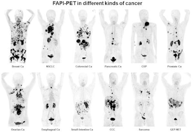 Ga-68 FAPI PET/CT shows 12 different tumor entities. Image courtesy of Kratochwil et al and SNMMI.
Should this paper be voted Scientific Paper of the Year by the Minnies expert panel, it would mark a twofer for this team of researchers from Germany: An image from the paper was named Image of the Year at this year's Society of Nuclear Medicine and Molecular Imaging (SNMMI) meeting.
In the study, a research team led by Dr. Clemens Kratochwil from University Hospital Heidelberg tested a new gallium-68 (Ga-68)-based radiotracer that acts as a fibroblast activation protein inhibitor (FAPI), which targets proteins that are overexpressed in cancer. Their goal is a radiopharmaceutical that will enable the clear visualization of tumors with high contrast.
What's more, researchers hope that Ga-68 FAPI could be used with PET/CT to visualize a wide range of tumors, such as breast, esophagus, lung, pancreatic, and head and neck cancers. Indeed, in the study they presented at SNMMI 2019, the researchers showed that Ga-68 FAPI demonstrated uptake in 12 types of cancerous tumors.
The researchers believe that Ga-68 FAPI could have advantages over the most widely used PET tracer, FDG, thanks to the shorter patient preparation required and a short radiotracer uptake time of just 10 minutes. Ga-68 FAPI could also be used for theranostic purposes if a therapeutic payload were attached to the radiotracer.
NELSON study shows CT screening for nodule volume management reduces lung cancer mortality by 26% in men. De Koning HJ et al, World Conference on Lung Cancer, September 2018. To learn more about this paper, click here.

Our second finalist for Best Scientific Paper is a landmark study that did for Europe what the National Lung Screening Trial (NLST) did for the U.S. -- show that CT lung cancer screening of high-risk smokers can significantly reduce mortality.
Only NELSON produced results even better than those of NLST, with a 26% mortality reduction in men compared with the 20% reduction in the NLST. (To be fair, the comparison isn't totally apples to apples, as the control group in NLST still got x-ray screening, while NELSON controls received no screening at all.)
NELSON was commissioned as a randomized, prospective study of CT lung cancer screening in 16,000 individuals in the Netherlands and Belgium over 10 years. Like NLST, the study was powered to definitively answer the question of whether CT lung screening should be rolled out on a population basis to high-risk people such as smokers.
Early NELSON results presented at the World Conference on Lung Cancer in September 2018 would seem to answer that question definitively in the affirmative. There were some subtle differences compared with NLST, the most important of which was the sharp mortality reduction that CT lung screening produced in women -- 61% compared with 26% for men. NLST's 20% reduction was for both men and women.
If NLST was a landmark study, NELSON has been just as important, if not more so, indicating that CT lung screening could be even more beneficial than previously thought.
Best New Radiology Device
Adaptive imaging receive (AIR) flexible MRI radiofrequency coils, GE Healthcare
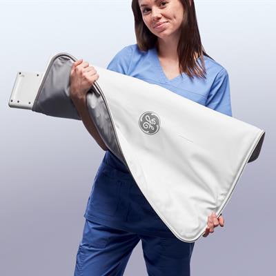 AIR radiofrequency coils. Image courtesy of GE Healthcare.
GE Healthcare is typically known for big iron, but the company's entry in this year's Minnies competition is light and flexible -- adaptive imaging receive (AIR) radiofrequency (RF) coils for GE's line of MRI systems.
The blanket-like coils are designed to address a nettling problem in MRI: Typical RF coils are bulky and awkward to use, and the time spent positioning them around a patient can slow down workflow in a busy MRI suite.
GE's AIR coils are 60% lighter than conventional coils and can be wrapped around patients for better image quality. The company has also developed special AIR Touch software that works with the coils -- the software recognizes patient anatomy and optimizes scan protocols, further reducing time required to set up patients.
First introduced at RSNA 2018, the AIR coils are available in a 30-channel anterior array coil, a 21-channel multipurpose large coil, and a 20-channel multipurpose medium coil. The coils will be available on GE's 1.5- and 3-tesla systems.
Elekta Unity MR radiation therapy system, Elekta
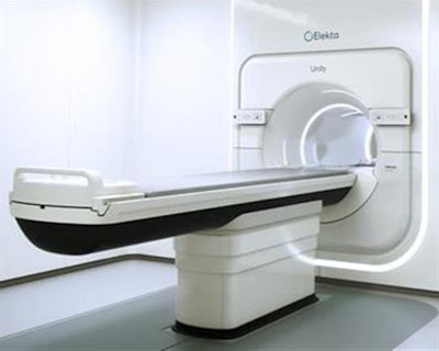 Elektra Unity. Image courtesy of Elekta.
Our second finalist in the Best New Radiology Device category represents a remarkable feat of engineering: the incorporation of an MRI system into a linear accelerator for MR-guided radiation therapy.
Unity integrates a 1.5-tesla MRI scanner with an advanced linear accelerator and software to deliver targeted radiation doses while simultaneously acquiring MR images for clinicians to visualize tumors and direct appropriate treatment. Unity was first displayed at the 2017 European Society for Radiotherapy and Oncology meeting, and it received U.S. Food and Drug Administration clearance in December 2018.
Elekta believes that Unity enables users to take advantage of MRI's ability to visualize soft tissue and employ it for guiding radiation treatments. In particular, the company is positioning Unity as a technology that can detect subtle changes in tumors between when reference images are acquired during treatment planning and when the treatment actually takes place.
Unity can potentially also provide functional MRI information on whether a tumor is responding to treatment, enabling radiation therapy to be made even more precise.
Best New Radiology Software
Aidoc algorithm for detection of pulmonary embolism, Aidoc
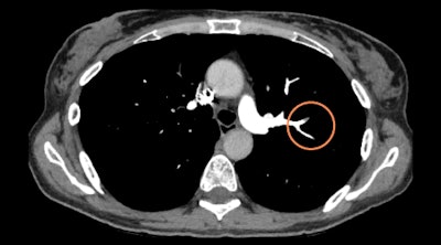 Aidoc's pulmonary embolism detection algorithm flags CT angiograms for urgent review. Image courtesy of Aidoc.
Israeli artificial intelligence (AI) software developer Aidoc's pulmonary embolism detection algorithm is designed to identify blood flow obstructions on pulmonary CT angiograms and flag critical exams for urgent review by radiologists.
Analyzing images right after they are acquired, the deep learning-based software can assist in managing pulmonary embolism cases without disrupting workflows and enable radiologists to quickly attend to time-sensitive and potentially life-threatening cases, according to the vendor. Researchers at the University Hospital of Basel in Switzerland highlighted the benefits of the software in a presentation at ECR 2019 in Vienna.
The algorithm received U.S. Food and Drug Administration (FDA) clearance in May.
PowerScribe One radiology reporting software, Nuance Communications
First unveiled at RSNA 2018, the next generation of Nuance Communications' PowerScribe radiology reporting platform features the integration of diagnostic and decision-support tools based on artificial intelligence (AI).
PowerScribe One harmonizes applications on the radiologist's desktop, automatically populates reports with structured data from integrated systems, and integrates AI image characterization directly into the reporting workflow, according to the vendor. The cloud-based platform also converts unstructured speech-to-text input into structured data. What's more, PowerScribe facilitates information sharing with care teams and across the clinical pathway, as well as enables standardization of reporting initiatives, the company said.
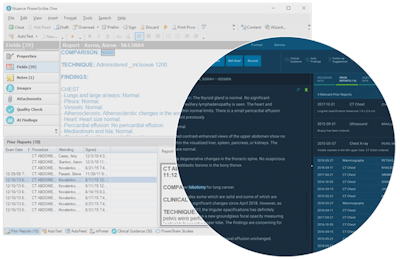
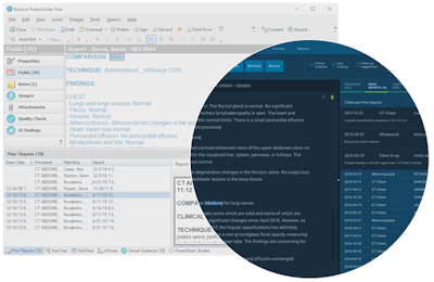 The PowerScribe One radiology reporting software integrates artificial intelligence-based diagnostic and decision-support tools. Image courtesy of Nuance Communications.
Best New Radiology Vendor
CureMetrix
There's no question that artificial intelligence is hot (indeed, the growth of AI is a finalist for the Most Significant News Event). So perhaps it's no surprise that a developer of AI software is also in the running for Best New Radiology Vendor.
CureMetrix has developed cmTriage, a software application designed to support radiologists interpreting mammograms -- but don't think of it as traditional computer-aided detection (CAD). Instead, cmTriage is designed to help radiologists triage their breast screening workload by customizing, sorting, and prioritizing their mammography worklist based on cases the software has determined may require immediate attention.
cmTriage gained U.S. Food and Drug Administration 510(k) clearance in March 2019 for use as a notification-only tool used in parallel with a radiologist's workflow to flag patients whose images might need more attention.
CureMetrix points out that cmTriage works with 2D full-field digital mammography equipment from any vendor. The software takes less than five minutes to process a study, so it doesn't slow down workflow.
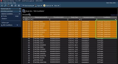 cmTriage uses the power of AI to triage mammograms for radiologist review. Image courtesy of CureMetrix.
United Imaging Healthcare
It's been a while since a new multimodality vendor appeared on the radiology scene, but United Imaging Healthcare is filling that role with a full line of MRI, CT, PET, digital radiography, and radiation therapy systems that are beginning to hit the market.
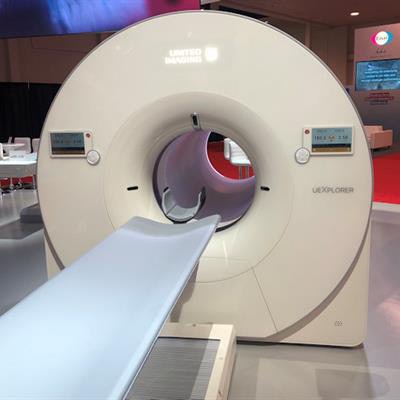 uExplorer PET system. Image courtesy of United Imaging Healthcare.
While United's product line spans the gamut of imaging, perhaps the company's crown jewel is uExplorer, a PET system that features a line of PET detectors along its gantry; the system is capable of acquiring whole-body images in a single bed position in 20 to 30 seconds. United believes that uExplorer makes possible the continuous tracking of radiopharmaceuticals as they are distributed in blood, organs, and tissue.
uExplorer was developed in collaboration with the University of California, Davis, and the first clinical system went into use at the university in August. The company has also begun installing other systems, such as the uMI 550 PET/CT scanner installed in June at Southwest X-Ray in El Paso, TX.
United's origins are in Shanghai, China, but the company is rapidly building up a U.S. presence and plans to manufacture system at its U.S. headquarters in Texas. The company now has more than 3,000 employees, over half of whom are involved in R&D, according to the company.
Best Educational Mobile App
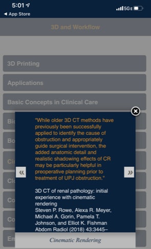 CTisus iPearls. Image courtesy of Dr. Elliot Fishman.
CTisus iPearls, Dr. Elliot Fishman (iOS)
A perennial contender in our Best Educational Mobile App category, Dr. Elliot Fishman of Johns Hopkins University has added another finalist award for another member of his CTisus family of apps, CTisus iPearls.
CTisus iPearls app features "pearls," facts or bits of knowledge that can help radiologists to make the right diagnosis or better understand an image.
The app includes 29 popular topics with pearls from the literature or the institution's imaging experiences, according to Fishman. Released in June, version 7.5 includes thousands of pearls.
CTisus iPearls, like other CTisus apps, is available for free.
Radiology Assistant 2.0, BestApps (iOS)
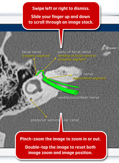 Radiology Assistant 2.0. Image courtesy of Dr. Woulter Veldhuis, PhD.
The defending champion in the Best Educational Mobile App category returns as a finalist for the 2019 Minnies. Radiology Assistant 2.0 features offline and interactive access to concise radiological expertise via peer-reviewed articles from expert radiologists around the world, according to Dr. Wouter Veldhuis, PhD, a radiologist at University Medical Center Utrecht in the Netherlands.
Radiology residents and fellows in 107 countries frequently consult the app during daily reading of clinical cases, he said. Interestingly, Radiology Assistant 2.0 is also popular with residents of other specialties, who use the app to read up on relevant topics, algorithms, or imaging appearances before visiting patients, consulting a radiologist, or participating on a multidisciplinary team, Wouter said.
Radiology Assistant 2.0 is free to download, but a subscription is required to access article categories. Contributions are used to support the Medical Action Myanmar medical aid organization.
Previous page | 1 | 2 | 3


