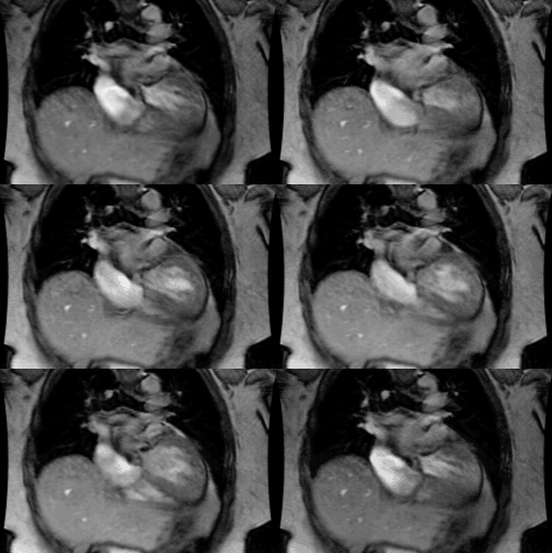
Mitral Valve Prolapse:
View cases of mitral valve prolapse
Clinical:
Mitral valve prolapse (MVP) is one of the most prevalent cardiac valvular anomalies affecting between 5 to 10% of the population and there is a strong hereditary component. The syndrome is secondary to a myxomatous degeneration of the mitral valve leaflets. Prolapse is now defined as more than 2 mm of left atrial displacement of the mitral leaflets from the line connecting the hinge points (i.e.: beyond the annular plane into the atrium) and a leaflet thickness of at least 5 mm in diastole [2,4] (although others indicate that valve thickening may or may not be present [3]). Although most patients are asymptomatic, the severity of the syndrome increases with age and up to 15% will develop progressive mitral regurge. Patients demonstrate a characteristic midsystolic "click" and mid to late systolic murmur, but this often does not occur until young adulthood. Complications include progressive mitral regurge (predominantly males, over age 50 years), infective endocarditis, and thromboemolic disease. Concurrent tricuspid valve prolapse may be seen in up to 20% of patients.
X-ray:
A body habitus characterized by a narrow anterior-posterior diameter, straight back, scoliosis, and a pectus excavatum or carinatum deformity may be noted (similar to that found in association with Marfans). Echocardiography is the best imaging modality for making the diagnosis of MVP, but the condition can also be identified by MRI.
REFERENCES:
(1) J Thorac Imag, 95, 10: p.1-25
(2) Society of Thoracic Radiology Annual Meeting 2000 Course Syllabus; Miller SW. Problem solving techniques for cardiac cases. 150-152
(3) AJR 2010; Shah RG, et al. Mitral valve prolapse: evaluation with ECG-gated cardiac CT angiography. 194: 579-584
(4) Radiographics 2010; Morris MF, et al. CT and MR imaging of the mitral valve: radiologic-pathologic correlation. 30: 1603-1620


