Case 2: Heavily calcified aortic valve
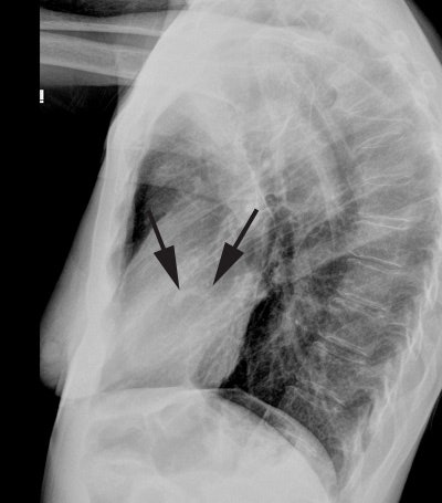
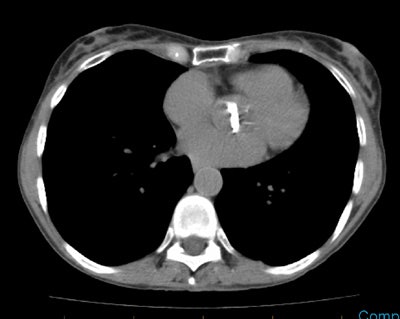
The lateral CXR below revealed dense calcifications in the region of the aortic valve (black arrows). When seen on plain film, the findings is very suggestive of aortic stenosis. CT imaging in this same patient also revealed very dense aortic valve calcifications.


