AJR Am J Roentgenol 1989 Oct;153(4):727-730
Chronic eosinophilic pneumonia: CT findings in six cases.
Mayo JR, Muller NL, Road J, Sisler J, Lillington G
Department of Radiology, University of British Columbia, Vancouver, Canada.
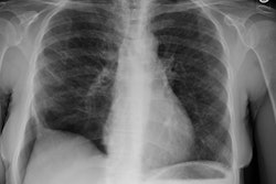
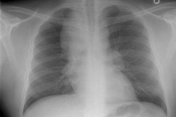
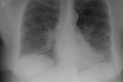
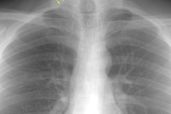
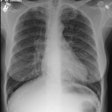
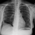
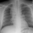
We reviewed the chest radiographs and CT scans in six patients with proved chronic eosinophilic pneumonia. In all patients, the chest radiographs showed patchy air-space consolidation, and in five of six cases, the consolidation was most marked in the middle and upper lung zones. In only one patient was the classic pattern of air-space consolidation that is confined to the outer third of the lungs readily apparent. In three patients, the consolidation appeared to be diffuse, although a slight peripheral predominance was present. In two patients, a peripheral predominance was difficult to appreciate, even in retrospect. The CT scans in all cases showed peripheral air-space consolidation. In addition, mediastinal adenopathy was identified on CT scans in three cases. This has not been described before in association with chronic eosinophilic pneumonia. A follow-up CT scan in one patient showed resolution of the adenopathy and marked improvement in the peripheral air-space disease within 2 weeks. We conclude that patients with chronic eosinophilic pneumonia show predominantly peripheral air-space consolidation on CT scans, even when this distribution is not readily apparent on the radiograph. CT may be helpful in the diagnosis when the clinical findings are suggestive, but the radiographic pattern is nonspecific.
PMID: 2773727, MUID: 89371259
