Koyama T, Ueda H, Togashi K, Umeoka S, Kataoka M, Nagai S.
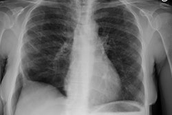
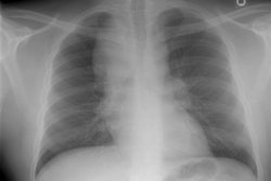
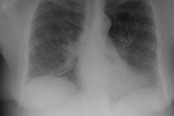
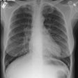
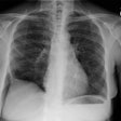
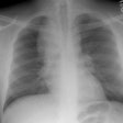
Sarcoidosis is a systemic disorder of unknown cause with a wide variety of clinical and radiologic manifestations. The diagnosis is usually made on the basis of these manifestations supported by histologic findings. Systemic manifestations (eg, Lofgren syndrome, Heerfordt syndrome) are commonly seen at clinical examination. Bilateral hilar lymphadenopathy is the most common radiologic finding-frequently with associated pulmonary infiltrates-and typically has a characteristic perivascular distribution at high-resolution chest computed tomography. Radiologic findings in the short tubular bones of the hands and feet and magnetic resonance imaging findings of nodular involvement of muscle are often sufficient to raise suspicion for sarcoidosis. In the liver, spleen, kidneys, and scrotum, coalescing granulomas form nodules whose imaging features may occasionally be nonspecific, although familiarity with the relevant clinical settings will be helpful in recognizing the presence of sarcoidosis. Radiologic recognition of cardiac and central nervous system involvement is also important because patients may be only mildly symptomatic. The clinical course and prognosis of sarcoidosis are highly variable, often correlating with the mode of onset. Familiarity with the clinical and radiologic features of sarcoidosis in various anatomic locations plays a crucial role in diagnosis and management. Copyright RSNA, 2004.

