J Thorac Imaging 1986 Mar;1(2):75-93
Patient work-up for bullectomy.
Gaensler EA, Jederlinic PJ, FitzGerald MX
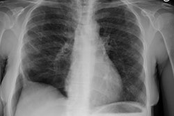
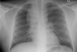
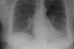
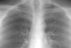
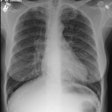
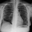
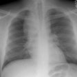
A localized area of hypertransradiance often leads to surgical referral. Among 608 cases, 115 were due to local lesions of airways, blood vessels, or parenchyma. Among the remaining 493 with bullae from diffuse emphysema, 21% underwent surgery. Good restoration of function occurred in patients with rapidly progressive dyspnea who did not have a bronchitic component, recurrent infections, or CO2 retention. Physiologically, preoperative findings suggestive of tension pneumothorax, including severe restriction, marked air trapping, and little ventilation/perfusion mismatch suggested good results. Favorable radiographic findings included well-defined, large air spaces without stigmata of diffuse emphysema, serial films showing rapid enlargement of bullae, and expiration films with good thoracic motion and obscuration of lung around bullae. Compressed but otherwise intact lung was best demonstrated by angiography and CT scans. Palliative bullectomy in severe diffuse emphysema sometimes had gratifying clinical results. Resection of small bullae never caused improvement. Localized giant bullae most often were associated with paraseptal or periacinar emphysema, and the best surgical results were obtained in this group.
PMID: 3599138, MUID: 87254357
