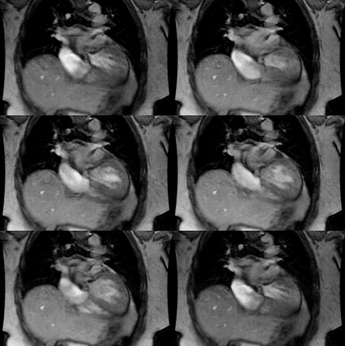
Tricuspid Valve Disease:
Clinical:
The tricuspid vale is a trileaflet structure (septal, anterior, and posterior cusps) and consists of three papillary muscles [3].Etiologies of tricuspid valve disease include: 1- Late stage rheumatic heart disease- may produce either stenosis (rheumatic heart disease is the most common cause of tricuspid stenosis [2]) or incompetence and is almost always associated with mitral valve disease); 2- Carcinoid syndrome (stenosis); and 3- Bacterial endocarditis (IV drug users). Clinically, patients have an enlarged pulsatile liver and increased jugular venous pressures. Doppler echocardiographic findings that are indicative of a hemodynamically significant stenosis include a mean pressure gradient of more than 5 mm Hg, an inflow time-velocity integral greater than 60 cm a pressure half-time greater than 190 msec, and a valve area by continuity equation of less than 1 cm2 [4]. On CXR there is right atrial enlargement.
Tricuspid insufficiency is most commonly secondary to dilatation of the RV and tricuspid annulus with streching of the leaflets- typically in response to elevated pulmonary arterial pressures [2].
The tricuspid valve is typically difficult to visualize by CT because of the relatively thin leaflet structure and contrast mixing artifacts [3].
REFERENCES:
(1) Radiologic Clinics of North America 1999; Lipton MJ, Coulden
R.
Valvular heart disease. 37(2): 319-339
(2) Radiographics 2009; Chen JJ, et al. CT angiography of the
cardiac valves: normal, diseased, and postoperative appearances.
29:
1393-1412
(3) J Cardiovasc Comput Tomogr 2012; Buttan Ak, et al. Evaluation
of
valvular heart disease by cardiac computed tomography assessment.
6:
381-392
(4) Radiographics 2015; Malik SB, et al. The right atrium: gateway to the heart- anatomic and pathologic imaging findings. 35: 14-31


