Sarcoidosis:
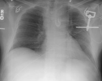
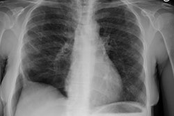
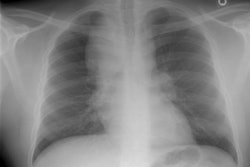
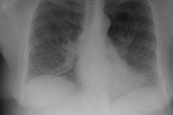
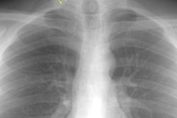
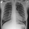
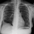
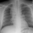
This is an interesting case of an active duty soldier that presented with complaints of shortness of breath. The CXR below demonstrates the presence of increased right paratracheal density, soft tissue fullness in the aortico-pulmonary window, and right hilar soft tissue fullness suggestive of mediastinal and hilar adenoapthy. No parenchymal abnormality was appreciated. Although the patient was suspected of having sarcoidosis, the patient's CT scan demonstrated the presence of an anterior mediastinal mass (best seen on the first image) associated with mediastinal and hilar adenopathy. There was a concern that the patient may have lymphoma, however, lung windows from the patients chest CT revealed very subtle, small nodules distributed within the upper lung zones bilaterally- a finding highly suggestive of sarcoid. The patient underwent transbronchial biopsy and the diagnosis of sarcoid was confirmed histologically.
