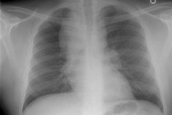
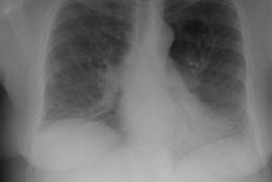
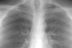
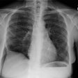
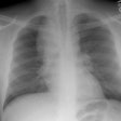
Earlier interstitial lung disease:
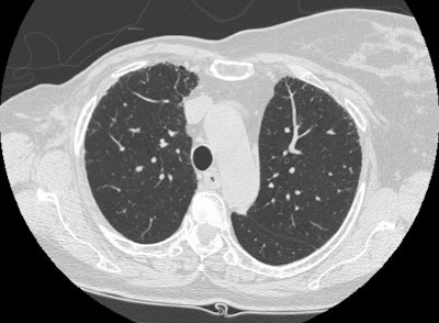
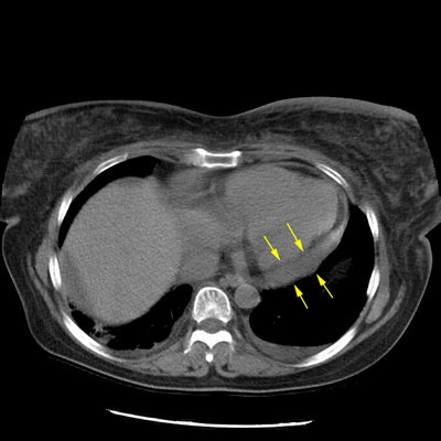
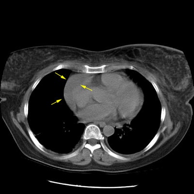
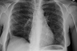
The case below is an example of interstitial lung disease at an earlier stage in a patient with a history of breast cancer (and a prior right mastectomy). There are patchy areas of fine subpleural "lace-like" intralobular interstitial thickening. Thickened interlobular septa are also seen, as is honeycombing- particularly in the posterior costophrenic sulci. The fibrosis associated with interstitial lung disease produces architectural distortion which is not seen in patients with lymphangetic metastases.
(Click on image to enlarge)
