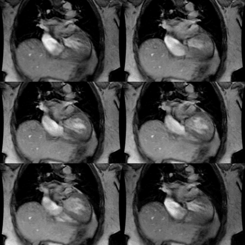
Lipomatous Hypertrophy of the Interatrial
Septum:
Clinical:
Lipomatous hypertrophy of the interatrial septum (LHAS) is characterized by the excessive deposition of fat (unencapsulated lipoblasts and mature fat cells [4]) in the interatrial septum and it is found in about 1% of patients at autopsy. LHIS is defined as the accumulation of adipose tissue within the septum exceeding 15mm and sparing the fossa ovalis [5]. The septum can exceed 2 cm in diameter [3]. It?s estimated prevalence is 2-8% by echocardiography and 2.8% by CT [5]. There is increasing prevalence associated with older age and a correlation with obesity and pulmonary emphysema [8]. There is also a higher prevalence in women [3].
In
most patients the condition is asymptomatic, however, rarely
an
association
with atrial arrhythmias,
obstructive
symptoms and
sudden death has been described [1,2].
X-ray:
On
computed tomography LHAS appears as a non-enhancing, smoothly
marginated, homogeneous, fat
density mass
(typically .20mm)
confined to the interatrial
septum- it
often has a
dumbbell configuration resulting from sparing of the fossa
ovalis [3,4]. Increased epicardial
fat and mediastinal lipomatosis
are frequently noted in association with LHAS.
PET imaging with FDG can show increased tracer uptake in the septum due to the presence of metabolically active brown fat in the abnormality [5,6]. Uptake can be seen in up to 82% of patients with LHIS- mean SUV 5.6 [7]. Fusion PET/CT helps to clarify the location of the uptake [7].
REFERENCES:
(2) AJR 2004; Tatli S, et al. MRI of atypical lipomatous hypertrophy of the interatrial septum. 182: 598-600
(3) Radiographics 2010; Kimura F, et al. Myocardial fat at cardiac imaging: how can we differentiate pathologic from physiologic fatty infiltration. 30: 1587-1602
(4) J Cardiovasc Comput Tomogr 2011; Rajiah P, et al. Computed tomography of cardiac and pericardiac masses. 5: 16-29
(5) J Cardiovasc
Comput Tomogr
2011; Rojas
CA, et al. Cardiac CT of non-shunt pathology of the interatrial
septum. 5: 93-100
(6) Radiographics 2011; James OG, et al.
Utility of FDG PET/CT in inflammatory cardiovascular disease.
31:
1271-1286
(7) Radiographics2011;
Maurer AH, et al. How to differentiate benign versus malignant
cardiac and paracardiac 18F
FDG
uptake at oncologic PET/CT. 31: 1287-1305
(8) Radiographics 2015; Malik SB, et al. The right atrium: gateway to the heart- anatomic and pathologic imaging findings. 35: 14-31


