Mitral Valve Prolapse:
(Case submitted by Dr. Scott Flamm)
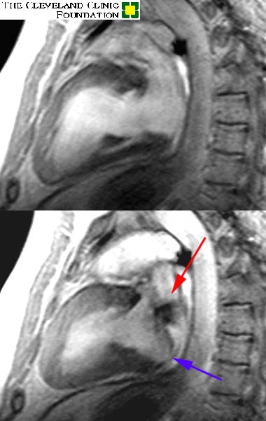
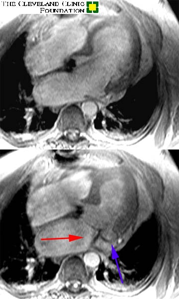
Image set #1 is a two chamber long axis view through the left ventricle and left atrium. Image set #2 is a four chamber view. The upper images are in diastole, while the lower are in systole. The red arrow demonstrates the turbulent jet of regurgitant blood flowing back into the left atrium. Regurgitation has occurred as a result of the 'prolapsing' posterior leaflet of the mitral valve noted by the blue arrow.
Image 1:

Image 2:


