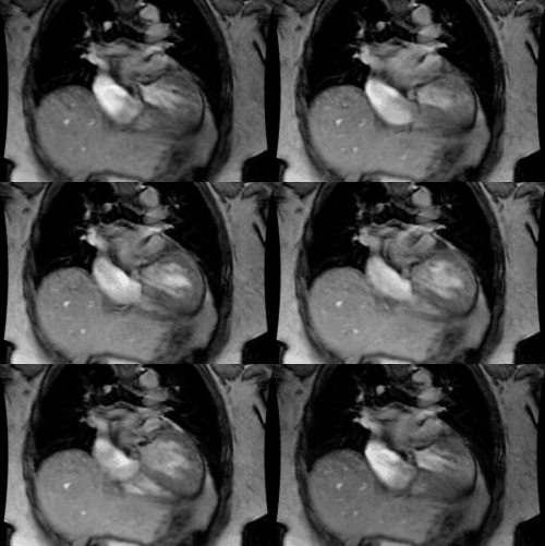
Polyspenia: Bilateral Left-sidedness- Left isomersim or Heterotaxy syndrome with polysplenia
Clinical:
Left isomerism or situs ambiguous with polysplenia is generally characterized by multiple spleens and midline or ambiguous location of the majority of the abdominal organs [3]. In the disorder, the structures normally on the left side of the body are seen on both sides, however, there is no fixed set of findings that are present in all cases. The main characteristic of left isomerism is bilateral left atrial appendages [6]. Because the sinus and atrioventricular nodes are right atrial structures, patients with left isomerism often have sinus node dysfunction or congenital heart block [6].Polyspenia is present in 55% of patients with left isomerism [6]. Polysplenia is associated with less severe cardiac malformations and patients may present at any age from infant to adult. There is a slight female predominance [4]. Polysplenia is characterized by the presence of multiple, small spleens, situs ambiguous, and absent gallbladder. The liver may have a central location in the upper abdomen and the left and right lobes are of equal size in about 25% of cases. Partial or complete failure of rotation of the intestinal tract is common. There is absence of the intrahepatic portion of the IVC with azygous or hemiazygous continuation in 50 to 85% of patients. Absence of the IVC is best identified as loss of the IVC shadow on the lateral CXR and azygous enlargement creates a paratracheal soft tissue density.
Bilateral bi-lobed lungs are present in two-thirds of the cases. Bilateral hyparterial bronchi (with the pulmonary artery crossing over the bronchus) are well demonstrated on CT with the pulmonary arteries seen posterior to the upper lobe bronchi.
Cardiac malformations are seen in 50-90% of cases, but they are often mild- such as partial anomalous pulmonary venous return, ASD (84%), or VSD (74%). Truncation of the pancreas is also commonly seen in adults [3]. Other findings include a pre-duodenal portal vein [5]. There are no Howell-Jolly bodies.
REFERENCES:
(1) J Thorac Imag, 1995, 10: p.43-57
(2) Radiographics 1999; Applegate KE, et al. Situs revisited: Imaging of the heterotaxy syndrome. 19: 837-852
(3) Radiographics 2002; Fulcher AS, Turner MA. Abdominal manifestations of situs anomalies in adults. 22: 1439-1456
(4) AJR 2009; Ghosh S, et al. Anomalies of visceroatrial situs.
193:
1107-1117
(5) J Cardiovasc Comput Tomogr 2012; Balan A, et al Atrial
isomerism: a pictorial review. 6: 127-136
(6) J Cardiovasc Comput Tomogr 2013; Wolla CD, et al. Cardiovascular manifestations of heterotaxy and related situs abnormalities assessed with CT angiography. 7: 408-416


