Alveolar Sarcoid:
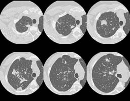
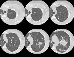
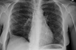
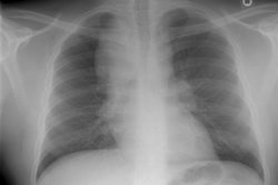
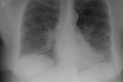
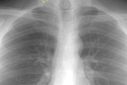
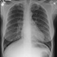
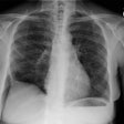
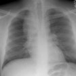
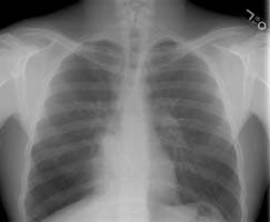
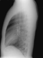
The female patient shown in the images below presented with complaints of dyspnea on exertion. Although many disease processes were entertained in the differential for the findings on her exams, a transbronchial biopsy was performed and a diagnosis of sarcoid was made histologically.
The CXR revealed bilateral, large nodular opacities with poorly defined
margins:
(Click small pictures to view larger radiographs)
A CT scan of the chest was performed and selected HRCT images were also
obtained:
(Click small pictures to view larger radiographs)
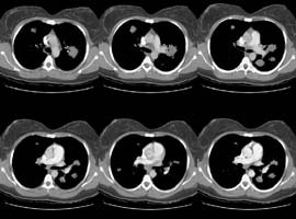
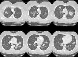
HRCT images:
(Click small pictures to view larger radiographs)
