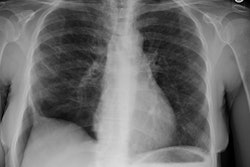
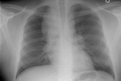
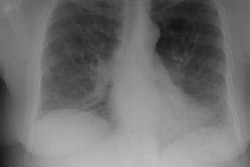
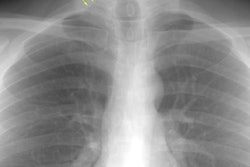
Plain radiographs and thoracic high-resolution CT in patients with ankylosing spondylitis.
Fenlon HM, Casserly I, Sant SM, Breatnach E
OBJECTIVE: The aims of this study were to identify the spectrum of abnormalities
seen on high-resolution CT in patients with ankylosing spondylitis and
to compare our findings with reports of plain film pulmonary manifestations
of the disease. SUBJECTS AND METHODS: We prospectively studied 26 patients
with documented ankylosing spondylitis. All patients underwent plain chest
radiography (posteroanterior and lateral views), thoracic helical CT, high-resolution
CT, and pulmonary function tests. RESULTS: High-resolution CT revealed
abnormalities in 18 patients (69%), whereas plain chest radiography revealed
abnormalities in four patients (15%). The most common abnormalities seen
on CT were interstitial lung disease (ILD) (n = 4), bronchial wall thickening
and bronchiectasis (n = 6), paraseptal emphysema (n = 3), mediastinal lymphadenopathy
(n = 3), tracheal dilatation (n = 2), and apical fibrosis (n = 2). CONCLUSION:
This study, which describes high-resolution CT findings in patients with
ankylosing spondylitis, reveals a spectrum of abnormalities unlike those
described in previous reports in which researchers used plain chest radiographs
as the sole imaging technique. In addition to apical fibrosis, high-resolution
CT revealed nonapical ILD, bronchiectasis, paraseptal emphysema, and tracheobronchomegaly.
Of these new findings, we believe that identification of ILD is the most
important. We suggest that nonapical ILD should be actively sought as an
explanation for pulmonary symptoms developing in patients with ankylosing
spondylitis. High-resolution CT should form an integral part of such workup.
