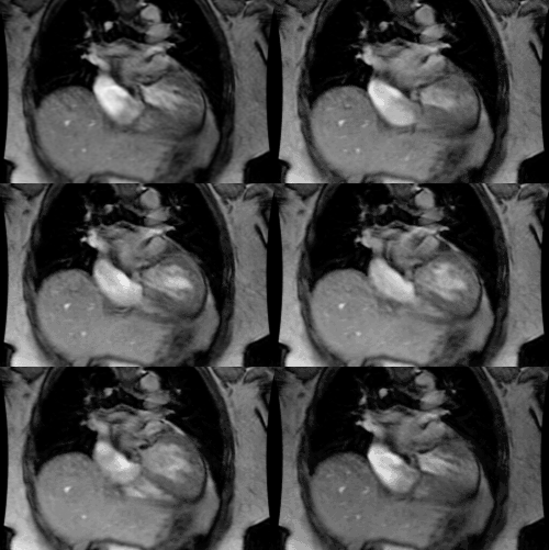
Partial Anomalous Pulmonary Venous Return:
Clinical:
Partial anomalous pulmonary venous return (PAPVR) is more common
than the total
variety with an overall incidence of about 0.5% [4]. PAPVR
results in an extra-cardiac left-to-right shunt, however, the
shunt is
usually hemodynamically insignificant [4].
Symptoms do not generally occur unless 50% or more of the
pulmonary
flow is shifted from
left to right (i.e.: a shunt ratio of pulmonary blood flow to
systemic
blood flow of greater than or equal to 1.5 [5]). Patients with
PAPVR
are usually older at presentation [5].
The right lung is more frequently affected than the left [4].
Drainage of the right upper lobe pulmonary vein directly into the
SVC
or right atrium
is the most common type of partial anomalous PAPVR [5] and this is
generally of no clinical
significance, although there is a high prevalence of an associated
sinus venosus ASD (located high in the septum near the SVC orifice
[4,6]).
Because this type of ASD is usually clinically silent, the
associated
PAPVR may be the clue leading to the diagnosis [6]. Some authors
suggest that right upper lobe PAPVR is associated with a sinus
venous ASD in 80-90% of pediatric patients, and 47% of adults [7].
Approximately one-third of patients with PAPVR have anomalous left pulmonary veins. In LUL PAPVR a verticle vein drains from the medial aspect of the left upper lobe into the left brachiocephalic vein. LUL PAPVR may be confused with a persistent left SVC- however, no connection to the coronary sinus will be seen and the left superior intercostal vein will not connect to the verticle vein associated with LUL PAPVR. Phase contrast MR images can also be used to demonstrate cephalic flow in the ventricle vein and caudal flow in the persistent SVC. [1]
Scimitar Syndrome (Pulmonary venolobar syndrome): Scimitar syndrome is due to a partial anomalous pulmonary venous return and patients are generally asymptomatic. The disorder is usually seen on the right side and can be identified as a "Scimitar" shaped vein crosses the lung. This vein usually drains to the IVC and the lung on the involved side is characteristically hypoplastic (as this is usually the right side there is a compensatory dextroposition of the heart). The pulmonary artery to the involved lung is also generally small or absent. Scimitar syndrome is associated with an accessory right hemidiaphragm (which can be identified as an increased retrosternal density). Ostium secundum atrial septal defects are usually present in association with PAPVR to the IVC [2].
REFERENCES:
(1) Radiographics 1997; MR imaging of congenital anomalies of the thoracic veins. 17: 595-608
(2) Seminars in Roentgenology 1989; Budorick NE, et al. The pulmonary veins. 24 (2) Apr: 127-149
(3) AJR 2000; Klecker RJ, et al. Vertical vein (Partial anomalous pulmonary venous drainage). 175: 869-870
(4) AJR 2004; Demos TC, et al. Venous anomalies of the thorax.
182:
1139-1150
(5) Radiographics 2012; Vyas HV, et al. MR imaging and CT
evaluation
of
congenital pulmonary vein abnormalities in neonates and infants.
32:
87-98
(6) Radiographics 2013; Porres DV, et al. Learning from the
pulmonary veins. 33: 999-1022
(7) Radiographics 2015; Sonavane SK, et al. Comprehensive imaging review of the superior vena cava. 35: 1873-1892


