Chronic Eosinophilic Pneumonia:
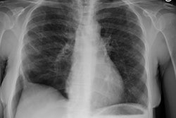
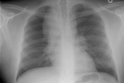
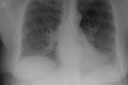
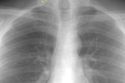
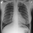
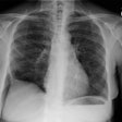
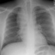
The patient shown in the radiographs below was a 53 year old male patient who presented with complaints of non-productive cough, progressive dyspnea, fever, and weight loss. Laboratory evaluation revealed an elevated erythrocyte sedimentation rate a mild leukocytosis with 20% eosinophils. Based upon the radiographic and lab findings, a diagnosis of chronic eosinophilic pneumonia was made. The patient responded to treatment with steroids.
Baseline chest radiograph:

Symptomatic chest radiograph demonstrated patchy peripheral areas of parenchymal consolidation:

Computed tomography again demonstrated patchy, bilateral, peripheral, non-segmental regions of consolidation which were sharply demarcated from adjacent normal lung. Cavitation was suggested in some regions of consolidation. Some of the opacities appeared streaky or band-like. Areas of ground glass opacification were also seen. Interlobular interstitial thickening and intralobular reticular opacities were noted in association with the areas of consolidation. Mediastinal adenopathy was identified and has been described in patients with chronic eosinophilic pneumonia. The appearance of CEP on computed tomography may be indistinguishable from bronchiolitis obliterans and organizing pneumonia (BOOP). Following institution of steroid treatment, the consolidations may begin to clear in as soon as two weeks- typically from the periphery.
Lung windows: 
Soft tissue windows: 
