Bronchocentric Granulomatosis:
View cases of bronchocentric granulomatosis
Clinical:
Bronchocentric granulomatosis is a rare disorder characterized by a necrotizing granulomatous inflammation of bronchial and bronchiolar epithelium/walls with chronic inflammatory changes in the adjacent lung parenchyma [3]. The disorder involves the bronchi (bronchocentric) and it is not actually a primary vasculitis (there are peribronchiolar and peribronchial necrotizing granulomas that do not invade the pulmonary arteries [6]). Necrotizing granulomatous lesions within the bronchioles produce small airway obstruction. Varying degrees of tissue eosinophilia (found in about one-third of patients [3]), chronic inflammatory changes occur in the surrounding peribronchiolar infiltrate, and manifestations of fungal hypersensitivity [4].Approximately one-third of cases are seen in asthmatic subjects. The disease tends to be most severe in patients with underlying asthma and these patients are usually males. These patients also have peripheral eosinophilia and often have serum precipitins positive to Aspergillus (suggesting a component of ABPA [3]). Other authors suggest that more than 50% of the cases are associated with asthma and allergic bronchopulmonary aspergillosis [6]. There is no evidence of renal or paranasal sinus disease.
Symptoms can be nonspecific and include fever, night sweats, cough, dyspnea, and pleuritic chest pain [4]. Affected patients without a clinical history of asthma tend to be older (average age 50 ), have no associated sex predilection, peripheral eosinophilia in only 50% of cases, and have a milder clinical course. Charcot-Leyden crystals (or aggregated eosinophilic granules) can be seen histologically [2].
The prognosis is good and symptoms can resolve spontaneously or with steroid administration [5].
X-ray:
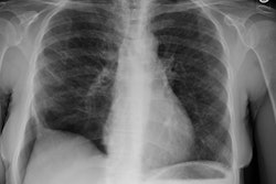
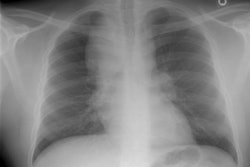
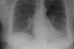
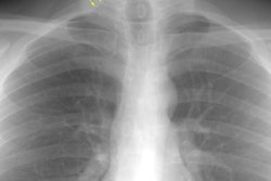
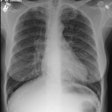
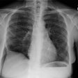
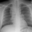
There is usually (60% of cases) a single area of mass-like opacification (representing a mass of necrotic tissue) associated with bronchiectatic, fibrotic, and/or cystic changes. Multiple nodules with ill-defined margins (centered on the airways [5]), areas of consolidation (30% of cases), mucoid impaction ("gloved-finger" branching shadows), reticulonodular infiltrates (10% of cases), and cavitation can be seen. The findings are unilateral in about 75% of cases and have an upper lung predominance in 60% of cases [3,4]. Hilar adenopathy and pleural involvement are rare [4].REFERENCES:
(1) Radiol Clin North Am 1991; Sept 29 (5): 973-982
(2) J Comput Assist Tomogr 1997; 21 (6): 920-930
(3) Radiographics 2007; Jeong YJ, et al. Eosinophilic lung
diseases: a clinical, radiologic, and pathologic overview. 27:
617-639
(4) Radiographics 2016; Katre RS, et al. Cardiopulmonary and
gastrointestinal manifestations of eosinophil-associated diseases
and idiopathic hypereosinophilic syndromes: multimodality imaging
approach. 36: 433-451
(5) AJR 2017; Bernheim A, McLoud T. A review of clinical and
imaging findings in eosinophilic lung diseases. 208: 1002-1010
(6) Radiographics 2020; Naeem M, et al. Noninfectious granulomatous diseases of the chest. 40: 1003-1019
