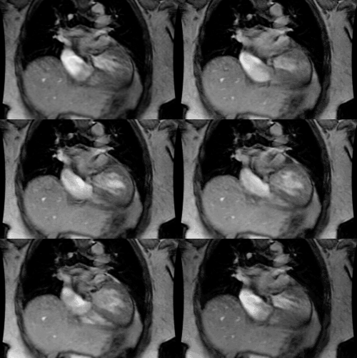
Pericardial Effusion:
View cases of pericardial effusion
Clinical:
Normally the pericardial sac contains 20-50 ml of fluid which serves to reduce friction between the beating heart and the pericardium. Etiologies of pericardial effusion include: Following myocardial infarction (Dressler's syndrome), following pericardiotomy, uremia, malignancy, infection (viral/TB), Kawasaki disease [2] and collagen vascular diseases such as SLE and Rheumatoid arthritis. AIDS is also a cause of pericarditis and pericardial effusion [3]. In fact, pericardial effusion is the most common cardiac finding in HIV patients and is associated with a 6 month mortality of 62% [3]. Pericardial effusions are particularly common in children with HIV [3].X-ray:
CXR: Pericardial effusion is rarely diagnosed on CXR until it is greater than 200 ml. On CXR the condition appears as a globular shaped, enlarged heart without significant pulmonary venous congestion. A "fat pad sign"- separation of the visceral and parietal fat layers of greater than 2 mm on the lateral exam- may sometimes be seen (about 15% of patients). Differential considerations for a big heart and normal vessels include: (3 P's and 2 M's) Pericardial effusion, Preload conditions (Aortic insufficiency, Mitral regurge), Prescription (Treated failure, Myocarditis/ Cardiomyopathy), Mimics (Anterior mediastinal masses which displace the heart such as thymic lesions or lymphoma), and Miscellaneous lesions (Thiamine deficiency [Beri-Beri]).CT: A pericardial space anterior the the RV greater than 5mm corresponds to a moderate effusion of 100-500 mL [4]. Findings of cardiac tamponade include flattening or inversion of the right atrial or RV wall, with compression of these chambers; inversion of the interventricular septum; distention of the SVC and IVC; and reflux of contrast into the azygous vein and IVC on contrast enhanced images [4].
MRI: MR imaging is useful in demonstrating small effusions even when echocardiography is negative- fluid may be asymmetrically distributed and not identified by echo. Uncomplicated effusions (transudative) usually demonstrate absent or low signal intensity on 1 images and high signal intensity on T2 images. Hemorrhagic or proteinaceous effusions will often have areas of medium or high signal intensity on T1 images. Cine images can be useful in accessing for chamber compression or diastolic collapse of the right sided heart chamber indicative of cardiac tamponade. Pericardial fluid typically has bright signal on GRE images.
REFERENCES:
(1) Magn Reson Imaging Clin N Am 1996; May 4(2): 237-251
(2) Radiology 1998; Chung CJ, et al. Kawasaki disease: A review.
208:
25-33
(3) AJR 2012; Lichtenberger JP, et al. What a differetial a virus
makes: a
practical approach to thoracic imaging findings in the context of HIV
infection- Part 2, extrapulmonary pulmonary findings, chronic lung
disease, and immune reconstitution syndrome. 198: 1305-1312
(4) Radiology 2013; Pericardial disease: value of CT and MR imaging. 267: 340-356


