J Thorac Imaging 1995;10(1):26-35. Tetralogy of Fallot: diagnostic imaging after palliative and corrective surgery.
Greenberg SB, Faerber EN, Balsara RK
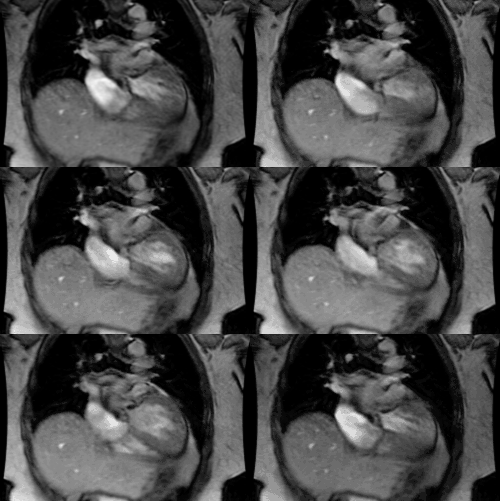
Tetralogy of Fallot was invariably fatal until the development of palliative and later corrective surgical procedures. The prognosis for children with tetralogy of Fallot continues to improve almost a half century after the earliest palliative surgical procedure was performed successfully. Imaging of the child and adult after surgery for tetralogy of Fallot remains an important challenge because surgical complications or limitations frequently require imaging for complete evaluation and further management of the patient. Traditional imaging by chest radiography and arteriography has been largely replaced by echocardiography and ultrafast and conventional CT, as well as magnetic resonance imaging. This article reviews those aspects of diagnostic imaging that are appropriate to study the postoperative chest in the child or adult with tetralogy of Fallot.
PMID: 7534360, MUID: 95198350


