Radiology 1997 Jun;203(3):715-719
Acute eosinophilic pneumonia: radiologic and clinical features.
King MA, Pope-Harman AL, Allen JN, Christoforidis GA, Christoforidis AJ
Department of Radiology, Ohio State University Hospitals, Columbus 43210, USA.
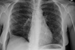
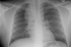
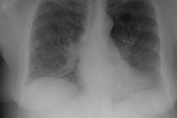
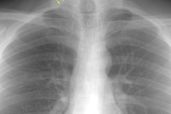
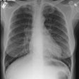
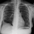
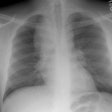
PURPOSE: To determine the radiographic and computed tomographic (CT) characteristics of acute eosinophilic pneumonia. MATERIALS AND METHODS: Twelve consecutive patients with acute eosinophilic pneumonia were included in the study. The diagnosis was based on clinical symptoms and results of bronchoalveolar lavage. Plain chest radiographs were obtained in all patients; CT scans were obtained in three patients. Two thoracic radiologists reviewed the radiographs and CT scans. RESULTS: Ten patients had bilateral areas of air-space opacity on images obtained at presentation; in seven of these patients, interstitial areas of opacity were also present. Two patients had bilateral interstitial areas of opacity and no areas of air-space opacity. Interlobular septal thickening and ground-glass attenuation were present on CT scans in two patients; patchy bilateral consolidation was present on CT scans in one patient. Pleural effusion was present on radiographs in seven patients (58%) and was bilateral in five. Pleural effusion was present at some point during the course of disease in all patients. In all patients, air-space disease markedly improved within 3 days of initiation of treatment with corticosteroids. CONCLUSION: Acute eosinophilic pneumonia should be considered as a possible diagnosis when a previously healthy person presents with acute respiratory failure of unknown origin.
PMID: 9169693, MUID: 97313089
