Cardiac Neoplasms:
Primary cardiac malignancies are very rare [11]. The incidence
of all primary cardiac tumors is estimated to be between 0.002-0.19% [14]. Approximately 75% of primary
cardiac tumors are benign and 25% are malignant [14,23]. The most
common benign cardiac tumors are myxomas (50%), papillary
elastomas (20%), lipomas (15-20%), and hemangiomas (5%) [23].
Malignant primary cardiac tumors are very rare and constitue only
10% of all primary cardiac tumors [23]. The majority (95%) of
malignant cardiac tumors are sarcomas or lymphomas (5%) or
pericardial mesotheliomas [23].
Secondary cardiac neoplasms (metastases) are much more common than primary lesions (about 20 to 40 times more common [23]) and can be identified in 10-20% of patients with an underlying malignancy at autopsy. Malignancies commonly associated with cardiac metastases include lung, breast, esophagus, kidney, sarcoma, and lymphoreticular malignancies [8,11,14,16]. Melanoma also has a propensity to metastasize to the heart [8]. The most common site of involvement is the pericardium with an associated malignant pericardial effusion (hemorrhagic or exudative) [23]. Cytologic studies are positive in 80-90% of patients with malignant pericardial effusions [8].
Fibroma:
- Clinical:
Cardiac fibromas is a very rare benign congenital neoplasm
that typically affects children, one-third of whom are under 1
year of age [6]. It is the second most common primary benign
cardiac neoplasm in children (after rhabdomyoma)
[16]. About 15% of cardiac fibromas
occur in adolescents and adults. The ratio of pediatric to adult
patients is 4:1, but there is no sex predilection [30]. Cardiac fibroma are almost always solitary and are
located in the ventricular myocardium- typically the left
ventricular free wall, the interventricular
septum, or the right ventricular free wall (in declining order) [1,14,28,30]. The central portion of the
tumor often has multiple areas of calcification and cystic
degeneration.
One third of affected patients are asymptomatic [6,26,28]. Common clinical findings include heart failure, arrhythmia, and sudden death [6]. Treatment is surgical excision for symtomatic patients as cardiac fibromas may remain stable for many years or regress [6]. However, other authors recommend resection even in asymptomatic patients due to the increased risk for sudden cardiac death [14] and lack of regression [26]. Recurrence following surgical excision is rare [6].
The lesion is associated with familial adenomatous polyposis syndromes (and its subtype Gardner syndrome) and Gorlin's (Gorlin-Goltz) syndrome (basal cell nevus syndrome [autosomal dominant] consisting of multiple nevoid basal cell carinomas of the skin, odontogenic keratocysts of the mandible, bifid ribs, and a tendency toward neoplastic growth in several organ systems [7]). However, less than 14% of patients with Gorlins syndrome have cardiac fibromas (some authors suggest only 3-5% of patients with Gorlins have fibromas [26]) [6]. Tumors associated with polyposis syndromes appear to occur more commonly in the atria [14,23,25].
- X-ray:
The most common radiographic finding is cardiomegaly
[6]. A focal bulge may be seen if the tumor involves the
ventricular free wall [6]. Tumor calcification can be seen in
about 25% of cases [25]- and this aids in differentiation from rhabdomyomas.
On cardiac echo, fibromas appear as a non-contractile
heterogeneous solid mass [26].
CT will reveal a large (mean diameter 5 cm [28]) homogeneous
mural mass (hemorrhage and necrosis are absent)-
it may be sharply marginated or
infiltrative [7,16]. Other authors suggest that the lesion appears
heterogeneous on CT [25]. Some authors suggest that central
calcification is a common feature on CT which can aid in
differentiation from a rhabdomyoma [26,28]. The lesion often does
not enhance significantly after contrast administration [7], but
enhancement has been reported [6,7]. An
associated pericardial effusion may be seen [6].
On MR, the lesions appear as a discrete, well defined homogeneous
mural mass or focal myocardial thickening. Other authors suggest a
heterogeneous appearance with variable areas of mildly hypo- or
hyperintense signal on T1 and T2 sequences [25]. The lesion is
typically isointense (or hyperintense or hypointense)
to myocardium on T1 images, and hypointense
to the myocardium on T2 images [6,26]. There is only slight or no
significant early enhancement following contrast administration
compared with the surrounding myocardium because of the minimal
vascular supply [14,23]. However, other authors report that the
lesion enhances heterogeneously [6,26]. Perhaps this is because
the lesion classically shows intense hyperenhancement on delayed
(7-10 minute) imaging [23,25,30].
Hemangioma:
- Clinical:
Cardiac hemangiomas are rare,
typically solitary tumors that account for 5-10% of benign cardiac
tumors [11,23] and fewer than 25% of cardiac hemangiomas occur in
children [26]. Most patients are asymptomatic [6]. The tumor can
arise from the endocardium,
myocardium, or epicardium [14]. The
tumor is classified as cavernous, capillary, or arteriovenous
microscopically [25]. They are usually found in the ventricles
(particularly in the lateral wall of the left ventricle and less
commonly in the anterior wall of the RV or the septum), although
they may occasionally be multifocal [14,16].
However, other authors have indicated that they usually arise from
the right atrium and have an intracavitary
component. The lesion is usually
asymptomatic [25].The most common clinical presentation
is dyspnea on exertion [11], but they
can also cause ventricular tachycardia, high-output heart failure,
hemorrhage, pericardial effusion, and cardiac tamponade [25].
Patients may also have hemangiomas in other sites such as the
skin, oral cavity, or bowel [1,25].
Cardiac hemangiomas can occur in the
setting of Kasabach-Merritt syndrome-
which is characterized by mltiple
systemic hemangiomas associated with
recurrent thrombocytopenia and consumptive coagulopathy
[6]. Surgical excision is the treatment of choice for symptomatic
lesions with an excellent long term prognosis [6,25]. Asymptomatic
hemangiomas do not require surgery as spontaneous regression has
been reported [25].
- X-ray:
At echocardiography the lesion appears hyperechoic
[6]. The lesion is usually heterogeneous with areas of
interspersed fat on non-contrast CT and enhances intensely with contrast [6]. Phleboliths
are characteristic of an hemangioma [16].
On MR, similar to hepatic hemangiomas, the lesion has iso- to intermediate intensity on T1 images and become hyperintense on T2 FSE images [6]. Although, other authors indicate that they appear of increased signal on T1 images due to slow blood flow [23]. Intense, inhomogeneous enhancement has been reported following gadolinium administration [11,14].
Lipoma:
- Clinical:
Cardiac lipomas are very rare, typically solitary benign neoplasms [12]. Despite this fact, lipomas are the second most common primary benign cardiac neoplasm [16]. Cardiac lipomas have no sex predilection and can occur at any age, but are more commonly found in adults [6]. Nearly all cardiac lipomas are epicardial lesions, but myocardial or endocardial lesions may also occur (50% being subendocardial, 25% myocardial, and 25% subepicardial [12]) [6]. However, other authors report that half of cardiac lipomas originate from the subendocardial layer and the other half from the subepicardial or myocardial layers and grow into the pricardial sac [16]. The most commonly reported locations for the lesion are the right atrium, the left ventricle, and the interatrial septum [12]. The lesion may be asymptomatic. Symptoms include cardiac murmur, arrhythmias due to involvement of the cardiac conduction system, or symptoms of cardiac compression [6,7]. Multiple cardiac lipomas have been described in association with tuberous sclerosis [12]. Surgery is the treatment of choice, but tumors which infiltrate the heart may not be resectable [6].
- X-ray:
The most common radiologic finding is cardiomegaly
[6]. CT and MR images can demonstrate the fatty nature of the
lesion.
|
The patient below presented with chest pain. CXR revealed an abnormal contour to the right heart border. Echocardiography suggested an atrial mass and this was confirmed on CT imaging. Note low attenuation areas within the periphery of the lesion of fat attenuation. |
|
|
|
MR imaging confirmed the presence of fat within the lesion (T1 and T1 fat sat images shown below). Despite the presence of soft tissue elements within the lesion, the tumor was found to be an intramyocardial lipoma at pathologic analysis. |
|
|
Lipomatous Hypertrophy of the Atrium:
- Clinical:
The condition is not a true neoplasm (unlike a lipoma), but rather a focal collection of mature adipose tissue. It is found in obese, female, and elderly patients. There may be an association with cardiac arrhythmias. It is defined as any deposition of fat in the interatrial septum at the level of the fossa ovalis which exceeds 2 cm in transverse dimension (the condition spares the fossa ovalis) [7]. The term "hypertrophy" is actually a misnomer as the condition is caused by an increase in the number of adipocytes, not hypertrophy [7]. Because of the presence of brown fat, this entity may show FDG uptake on PET imaging [14].
Lymphoma (Primary Cardiac Lymphoma):
- Clinical:
Primary cardiac lymphoma is very rare - accounting for 1.3% of
primary cardiac tumors and less than 1% of extranodal lymphomas
[19,20]. The lesion is typically an aggressive non-Hodgkins B-cell type (diffuse large cell
lympphoma) [6,20]. To be considered a primary cardiac lymphoma,
these tumors involve only the heart or pericardium at the time of
diagnosis, with no evidence of extracardiac
lymphoma [6]. Immunocompromised patients, particularly HIV or
organ transplant, account for approximately 50% of cases [29]. The
lesion is seen with increased frequency in immunocompromised
patients with HIV due to Ebstein-Barr virus associated
lymphoproliferative disorders [6,16].
In HIV patients, a large pericardial effusion should raise
suspicion for a primary effusion lymphoma [24]. However, even in
HIV and organ transplant patients, it still represents less than
5% of all lymphomas in these patients [20].
Patients present with unresponsive, rapidly progressive heart
failure, dyspnea, arrhythmias, chest pain, and SVC syndrome. The
prognosis is poor and there is no evidence that surgery improves
prognosis [6]. Left ventricular involvement is associated with a
worse prognosis [19].
The lesion most commonly involves the right heart (particularly
the right atrium followed by the right ventricle) and is commonly
(75% of cases) multicentric with frequent involvement of more than
one chamber [11,25]. Pericardial thickening/involvement or
effusion are a common early feature of the disease [20,23].
Infiltration of atrial or ventricular walls with extension along
the epicardial surfaces encasing adjacent structures (including
the coronary arteries and aortic root) is also a notable feature
[20].
Lymphomas generally lack regions of central necrosis and
hemorrhage and are typically homogeneous [23]. On CT, the lesion
is often isoattenuating to hypoattenuating relative to myocardium
and demonstrates heterogeneous enhancement [20]. The lesion
usually appears homogeneous and isointense on T1 and T2 images
[23].
- X-ray:
The CXR usually demonstrates cardiac enlargement and evidence of heart failure. CT demonstrates a mass that is hypo- to isodense to myocardium with heterogeneous enhancement. There is often an associated large pericardial effusion [14]. In fact, a pericardial effusion may be the only finding [16]. The tumor can appear iso or hypointense to myocardium on both T1 and T2 FSE sequences and appear poorly marginated and heterogeneous following contrast administration [14].
Malignant Cardiac
Sarcomas:
- Clinical:
About 25% of primary cardiac tumors are malignant (most often secondary neoplasms related to metastases) [23]. The majority of primary cardiac malignancies are sarcomas (sarcoma is the most common primary cardiac malignancy [16]). Angiosarcoma (37-40% of cases) is the most common primary cardiac malignancy in an adult [11]), followed by undifferentiated sarcoma (24% of cases), leiomyosarcoma (8-9% of cases), rhabdomyosarcoma (more prevalent in children [11]), and malignant fibrous histiocytoma (11-24% of cases). Primary cardiac sarcomas affect adults and are extremely rare in infants and children [6]. Dyspnea is the most common presenting symptom [6]. Nearly 80% of patients with cardiac sarcomas have distant mets at presentation. Prognosis is uniformly poor.
Angiosarcoma: Cardiac angiosarcoma is the most common cardiac
sarcoma [4,6]. Patients generally
present between the ages of 20-50 years (middle aged men [14]).
They most commonly occur in the right atrium (80% of cases [6])
and produce valvular obstruction and
right ventricular failure. Pericardial involvement is common and
leads to the presence of a hemorrhagic pericardial effusion [4,6,23,24]. Multiple lesions occur in 60% of
patients [16]. Metastases occur in 66-89% of cases at the time of
presentation [6], most commonly to the lung [11]. Prognosis is
poor [4].
The typical CT findings of angiosarcoma include an irregular, nodular, broad-based right atrial mass with heterogeneous enhancement and pericardial infiltration (thickening or effusion) [16]. On MRI the tumor is typically heterogeneous with low signal intensity on T1 and increased intensity on T2 FSE images [14]. Areas of high signal are seen due to necrosis or blood filled spaces/hemorrhage within the tumor. A "cauliflower" of papillary appearance has also been reported. There is heterogeneous enhancement of the mass following contrast administration [10] with marked surface enhancement (a "sun ray" appearance has also been described [11]). Because of the tendency of the tumor to involve the pericardium, pericardial thickening or nodularity, or a hemorrhagic pericardial effusion may also be detected. [2,11]
Malignant Fibrous Histiocytoma: Although rare for the tumor to occur in the heart, it is the next most common cardiac sarcoma. It may occur more commonly in women. The lesion commonly arises from the posterior or septal wall of the left atrium. It can obstruct atrial filling and outflow leading to left sided heart failure. [2]
Undifferentiated sarcoma: Assount for about 1/3 of all cardiac sarcomas and are the second most common primary cardiac malignancy in adults [23]. The tumor can occur at any age, but patients are typically in their 4th or 5th decade, and the lesion has a propensity to arise in the left atrium (81% of cases) [4,16,23]. The tumor typically appears isointense compared to the myocardium on FSE images [14].
Leiomyosarcoma: Uncommon cardiac tumor (1% of all primary cardiac tumors [11]) with a predilection for the posterior wall of the left atrium (and invasion of the pulmonary veins is frequently seen) [14]. They usually affect patients during the 4th decade [14]. The most common presenting symptom is dyspnea and cardiac failure from mitral obstructive symptoms [11]. The lesion can be multiple in up to 30% of cases [4,11] Prognosis is poor. On MR the lesion is usually sessile, lobulated and appears hypointense to myocardium on T1 images, hyperintense on T2, and demonstrates marked contrast enhancement [11].
Fibrosarcoma: Fibrosarcoma tends to occur in the left atrium and usually amnifests as CHF [14].
Extraskeletal cardiac osteosarcoma: Account for 3-9% of cardiac sarcomas [16]. The tumor is usually centered in the left atrium and patients present with pulmonary congestion [14]. The lesion commonly contains large foci of calcification [14].
Mesothelioma: Pericardial mesothelioma
- Clinical:
Malignant pericardial mesothelioma is an extremely rare tumor, however, it is the most common primary malignancy of the pericardium [24]. The tumor is more common in men and can occur at any age, but is seen most often in the 5th through 7th decades with a mean age of 46 years at presentation [6,24]. Patients present with chest pain, dyspnea, cough, and palpitations [6]. Although there appears to be a relationship to asbestos exposure, a definite association has not been established due to the rarity of this lesion [6]. Surgery combined with radiation therapy may provide some palliation, but the prognosis is extremely poor with an average survival between 6 weeks to 15 months [6,24]. The tumor usually forms multiple coalescing masses that envelop the pericardial space [11]. Metastatic disease is found in approximately 50% of cases, most commonly to local mediastinal nodes and the lungs [24]. On CT there is nodular, irregular, diffuse pericardial thickening and a pericardial effusion [6].
Myxoma:
- Clinical:
Primary cardiac tumors are uncommon and approximately 75% of primary cardiac tumors are benign. Myxomas account for nearly half of the benign cardiac tumors encountered and are therefore the most common primary cardiac tumor [9,22]. It is a benign neoplasm which arises most commonly from the interatrial septum in the region of the fossa ovalis (78% of cases [16])- extending into the left atrium (60% to 75%) more commonly than the right atrium (20% to 28%) [14]. Ventricular myxomas are uncommon (about 5% of cases) [23]. Myxomas can present at any age (mean, 50 years), but are most common in the 4th to 7th decades and there is a female predominance (1.7:1 to 4:1) [6,23]. The tumor rarely occurs in children [25]. Left atrial lesions may prolapse through the mitral valve and cause atrial obstruction and hemodynamic alterations with dyspnea or syncope. The signs and symptoms are typically indistinguishable from mitral valve disease. Constitutional symptoms such as weight loss, fever, fatigue, and arthralgias are seen in about one-third of patients [6] and may be related to an autoimmune reaction initiated by the tumor [9]. Peripheral emboli or tumor emboli may also occur in 22% to 60% of patients (particularly with myxomas with irregular surfaces which have a tendency to induce thrombi formation [22])- most commonly to the brain (resulting in stroke), kidneys, or peripheral arteries (resulting in limb ischemia or infarction). Right sided myxomas will embolize to the lungs. About 20% of patients are asymptomatic [6]. Cardiac arrhythmias occur in 20% of affected patients [6]. Treatment is surgical excision with an excellent long term prognosis and low risk of recurrence. [5,6]
Cardiac myxomas are most commonly a sporadic lesion (93% of cases). About 94% are single, and the estimated risk of a second myxoma developing following surgical resection is 1-3% [6].
About 7% of cardiac myxomas exhibit atypical biologic behavior, including multicentricity, atypical location (in cardiac chambers other than the left atrium), recurrence after surgical excision (12-22%), and an association with unusual conditions such as Carney complex [6]. These patients are usually younger (mean age 28 years), more commonly male, and exhibit a familial predisposition for cardiac myxomas [6]. The lesions are associated with a higher risk of recurrence (12-22% of cases) [9]. The Carney complex is an autosomal-dominant, inherited disorder in which cardiac myxomas occur in association with cutaneous and mammary myxomas (myxoid fibroadenomas [9]), spotty skin pigmentations, endocrine overactivity (Cushing syndrome, sexual precocity, and acromegaly), psammomatous melanotic schwannoma, primary pigmented nodular adrenal disease, and testicular neoplasms (particularly Sertoli cell tumor) [6]. Cardiac myxomas occur in about two-thirds of patients with Carney complex [6], often at a younger age [16]. The myxomas can also occur outside the left atrium and have a higher rate of recurrence [16].
- X-ray:
The CXR can be normal in up to 37% of cases [9]. Radiographically visible calcification is present in up to 20% of cases (more common with right atrial lesions- up to half of right atrial tumors can have visible calcification on CXR [9]) [6]. Patients with left atrial tumors may exhibit findings similar to mitral valve disease with left atrial enlargement (for left atrial tumors). Pleural effusion in up to 15% of cases [9].
On CT the lesion is usually a well-defined, lobulated, intracavitary cardiac mass [6]. The lesion usually arises from a narrow stalk- most often from the fossa ovalis (this differs from thrombi that most commonly originate from the atrial appendage) [13]. The lesion is usually of lower density than unopacified blood on non-contrast images (HU 43 +/- 14 [17]), but the lesion is best identified after contrast administration [6]. The lesion is commonly heterogeneous reflecting hemorrhage, necrosis, fibrosis, and calcification [6] and is of lower density than the myocardium [9].
On MR the tumor is usually heterogeneous [9] and has a signal intensity similar to cardiac
muscle on T1-images [6] and has increased signal on T2 FSE images
[7]. Internally, myxomas may contain regions of necrosis,
fibrosis, hemorrhage, and calcifcation [23]. Following gadolinium
administration the lesion enhances heterogeneously [6]. Myxomatous components of the lesion appear
of low signal on T1 and high signal on T2 images [6].
Calcification will be of low signal on T1 and T2 images [6]. The
tumor appears of low signal intensity on GRE cine images and these
images can be used to demonstrate tumor motion and prolapse across the atrioventricular
valve [6].
On PET imaging, cardiac myxomas generally demonstrate very
low-grade or no significant FDG activity [18].
|
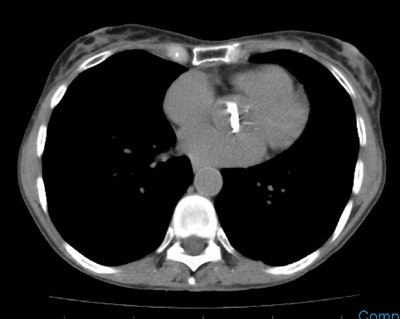
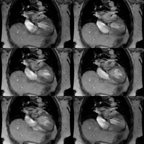
Right atrial myxoma: The patient below presented with pulmonary infiltrates and fevers. Chest CT was performed and demonstrated a possible lesion in the right atrial appendage. A follow-up cardiac MR confirmed a solid mass in the right atrial appendage (white arrows). The mass was surgically resected and found to be a myxoma. |
|
|
Papillary Fibroelastoma:
- Clinical:
Papillary fibroelastomas are benign endocardial papillomas that account for 10% of cardiac neoplasms and predominantly affect the cardiac valves (90% of lesions occur on cardiac valves) [7,11,16]. It is a rare lesion, but it is the second most common benign cardiac neoplasm [6,22] (however, other authors report that lipomas are the second most common benign neoplasm [16]). Most lesions are solitary and small (10-20mm) [16]. The lesion is characterized by avascular papillary fronds of connective tissue covered by a single layer of hypertrophied endothelium, with stroma of central amorphous fibrous core and outer loose connective tissue [16].
The lesion is more common on the aortic (29-44%) and mitral (25-35%) valves, than on the tricuspid (15-17%) and pulmonary (8-13%) valves [7,21,22]. About 16% of lesions arise from non-valvular surfaces (endothelial surface of the LV apex, the chordae tendineae, or LV outflow tract) [7,16,22]. Men and women are affected equally with a mean age at presentation of 60 years (peak incidence is in the 7th and 8th decades of life [21]) [6]. Most lesions are asymptomatic and are found incidentally at autopsy, cardiac surgery, or cardiac catheterization [6,7]. Symptomatic patients present as a result of systemic embolization of the tumor or thrombi on the tumor surface and may complain of chest pain, TIA's or stroke, dyspnea, or sudden death (due to coronary artery ostia occlusion) [14]. The embolic fragments may arise from the tumor itself or from platelet/fibrin clots on the lesion surface [6,16]. Treatment is surgical excision and recurrence has not been reported [6].
Differential considerations would include a vegetation or thrombus [11]. Vegetations are usually associated with destruction of the valve leaflets and valvular incompetence [11]. Myxomas can also occur on valves, but they are generally larger [11].
- X-ray:
Most lesions are imaged with echocardiography which reveals a small (under 1-1.5 cm), mobile, pedunculated, homogeneous valvular or endocardial mass which flutters or prolapses with cardiac motion [6]. The lesion is usually not evident on routine CT or MR imaging [7]. If seen, they are usually of intermediate or isointense signal intensity on T1 images and hyperintense on T2 FSE images [14,22]. The lesion can be seen on cine gradient images where it appears as a hypointense mobile mass with turbulent flow surrounding it [11,14,22].
Paraganglioma (Chemodectoma):
- Clinical:
Cardiac paragangliomas are extremely rare neoplasms that arise from intrinsic cardiac paraganglial (chromaffin) cells which are predominantly located in the atria (LA posterior wall or roof and less commonly the interatrial septum), AV groove, or near the roots of the great vessels [6,15,16]. A chemodectoma is a non-functioning paraganglioma [16]. Most lesions have been reported in adults (mean age 40 years) [6]. The majority of reported lesions have been catecholamine-producing tumors. Up to one-half of affected patients present with symptoms of catecholamine excess [15]- hypertension, palpitations, and flushing. Other clinical manifestations can result from mass effects of the tumor including invasion of the conducting system, compression of coronary blood flow, pericardial involvement, or disruption of valvular function [15]. Lab analysis will reveal elevated levels of urinary norepinephrine and vanillylmandelic acid [6]. Up to 20% of affected patients will have associated paragangliomas in other locations. Osseous metastases are found in 5% of cases. Surgery is the treatment of choice and it is often successful. A hypertensive crisis is a potential complication of surgical resection [6].
- X-ray:
Chest radiographs will demonstrate a middle mediastinal mass that splays the carina and may simulate left atrial enlargement [6]. Prior to CT scanning patients should be premedicated with alpha and beta blockers to prevent a hypertensive crisis [6]. CT will demonstrate a markedly enhancing mass adherent to or involving the left atrium (less commonly the mass can be located anterior to the aortic root). The lesion is typically located in the roof of the left atrium. Unlike myxomas, the lesion often has a broad base attachment [7]. About 50% of lesion have areas of central low attenuation due to necrosis. The lesions may contain calcification [6]. The lesion demonstrates intense, heterogeneous contrast enhancement [16]. The lesion may be missed on unenhanced or poorly enhanced scans as the mass may appear isodense to adjacent cardiovascular structures [6]. MR will demonstrate a low signal mass on T1 images which is very hyperintense on T2 images [6,7].
Rhabdomyoma/Rhabdomyosarcoma:
- Clinical:
Rhabdoid tumors arise from striated
muscle. Rhabdomyoma (a hamartoma of the
myocardium) is the most common primary pediatric cardiac
tumor- accounting for up to 90% of cardiac tumors in infants and
children [6,25]. The lesion is usually found in patients under 1 year of age [6]. Presentation in adults in rare [16]. The
incidence of tuberous sclerosis in patients with cardiac
rhabdomyomas is between 60-80%, and 47-67% of patients with
tuberous sclerosis have rhabdomyomas [11,23,25,26,27]. The tumors
are frequently multiple (60-90% of cases and multiplicity has an
even higher association with tuberous sclerosis [11,26]- unlike
fibromas which are usually solitary [23]).
The lesion most commonly arises intramurally in the ventricular myocardium (with equal frequency on the left or right [16]) and interventricular septum. Atrial wall involvement is much less common. Most patients are asymptomatic, but the tumor may produce obstruction to blood flow, murmur, arrhythmia, heart failure, and in utero fetal hydrops or still birth [25]. A prenatal diagnosis of cardiac rhabdomyoma confers a 75-80% probability for tuberous sclerosis [27]. Spontaneous partial or complete tumor regression may occur in fetal life and particularly in patients less than 4 years of age (in infants, 70% of tumors regress before the age of 4 years [27]), and surgical resection is not required unless there are clinical symptoms (such as dysrhythmias or heart failure/outflow obstruction) [14,25]. The prognosis is good, and management is conservative in the absence of life threatening symptoms.
Rhabdomyosarcomas account for about 20% of cardiac sarcomas and are the most common cardiac malignancy in infants and children [4,11]. There are two histologic subtypes- embryonal, which is more common, and pleomorphic [25]. The pleomorphic subtype is uncommon, but is more frequent in adults [11]. There is a slight male predilection [4]. Congestive heart failure is a common presenting symptom [11]. The lesion is often mutliple and can invade the pericardium [4]. The lesion is also more likely to involve the cardiac valves unlike other primary cardiac sarcomas [11,23]. On CT the lesion appears as a smooth or irregular low attenuation mass in the cardiac chamber. The lesion is isointense to myocardium on T1 images and enhancement is generally homogeneous, although areas of low signal can be seen in association with central necrosis [11]. The lesion can have homogeneous or heterogeneous signal on MR images [4].
- X-ray:
Echocardiography: Rhabdomyomas
appear as a solid echogenic mass that
may have an intracavitary component.
When the lesions are small and multiple and diffuse myocardial
thickening is the predominant finding [6].
On MR imaging, the tumor typically appears homogeneously iso- to marginally hyperintense
to cardiac muscle on T1 images and a slightly increased signal
intensity on T2 images [6,11]. Purely
intramural lesions may not be identified on MR imaging, although
they may be seen following contrast enhancement and appear hypointense to normal myocardium (i.e.- the lesion shows minimal or no
enhancement on first pass perfusion imaging), but becomes
isointense on late gadolinium enhanced sequences [11,14,25].
On MR, rhabdomyosarcomas appear as a large infiltrative mass with
irregular margins and central necrosis or cavitation [25]. The
lesion is isointense on T1, hyperintense on T2, and generally
demonstrate heterogeneous contrast enhancement due to areas of
necrosis [23,25]. There is peripheral hyperintensity on late
gadolinium enhanced sequences [25].
Teratoma:
- Clinical:
Teratomas are typically benign solitary, cystic, intra-pericardial lesions. The lesion typically affects infants and children, but is extremely rare [6]. It is usually extra-cardiac located in the pericardium [25]. They generally arise from the base of the heart near the origin of the great vessels and rarely involve the myocardium [25]. Patients usually present with respiratory distress and cyanosis secondary to cardiac tamponade and compression of right sided vascular structures. If patients present with tamponade an emergent pericardiocentesis is required. Treatment of the lesion is then surgical with a good prognosis [6].
- X-ray:
The lesion can be seen on prenatal US which demonstrates an intrapericardial multilocular
cystic mass associated with pericardial effusion and findings of
fetal hydrops [6]. Chest radiographs
demonstrate an enlarged cardiomediastinal
silhouette [6]. The presence of a formed tooth can confirm the
diagnosis if visible radiographically.
Echocardiography will reveal an intrapericardial
heterogeneous and complex multilocular
cystic mass, typically positioned on the right side of the heart
[6]. MR imaging will usually demonstrate a large mass of
heterogeneous signal intensity.
Thrombus:
- Clinical:
Thrombus represents a cardiac pseudotumor and is one of the most
common causes of an intracardiac mass [23].
- X-ray:
The T1 and T2 characteristics vary depending on the age of the
thrombus [23]. An acute thrombus usually has intermediate signal
on T1 and T2 because the hemglobin content is still in the
oxygenated state [23]. In subacuute thrombus, the hemoglobin has
been metabolized to methemoglobin which produces a lower signal on
T1, but high signal on T2 due to the high water content from lysed
red blood cells [23]. Over a greater time period, the thrombus
becomes water depleted and the cellular debris containing
methemoglobin is replaced by fibrous tissue and produces lower
signal on T1 and T2 images [23].
Because thrombus is avascular it does not enhance on post
contrast imaging [23]. rarely, very chronic, organized thrombus
may enhace peripherally due to its fibrous content [23].
REFERENCES:
(1) Radiographics 1997; Pediatric case of the day. Cardiac rhabdomyoma. 17: 805-807 (No abstract available)
(2) Radiographics 1997; 17: 145-153
(3) Magn Reson Imaging Clin N Am 1996; May 4(2): 237-251 (p. 248-250)
(4) Radiographics 1999; Araoz PA, et al. CT and MR imaging of primary cardiac malignancies. 19: 1421-1434
(5) Magn Reson Imaging Clin N Am 1996; May 4(2): 237-251 (p.245-47)
(6) Radiographics 2000; Grebenc ML, et al. Primary cardiac and pericardial neoplasms: Radiologic-pathologic correlation. 20: 1073-1103
(7) Radiographics 2000; Araoz PA, et al. CT and MR imaging of benign primary cardiac neoplasms with echocardiographic correlation. 20: 1303-1319
(8) Radiographics 2001; Chiles C, et al. Metastatic involvement of the heart and pericardium: CT and MR imaging. 21: 439-449
(9) Radiographics 2002; Grebenc ML, et al. Cardiac myxoma: imaging features in 83 patients. 22: 673-689
(10) Radiographics 2003; Best AK, et al. Cardiac angiosarcoma. 23: S141-S145
(11) Radiographics 2005; Sparrow PF, et al. MR imaging of cardiac tumors. 25: 1255-1276
(12) J Nucl Cardiol 2008; Ruggiero NJ, et al. Myocardial perfusion defect caused by intramyocardial lipoma. 15: 286-289
(13) AJR 2009; Scheffel H, et al. Atrial myxomas and thrombi: comparison of imaging features on CT. 192: 639-645
(14) AJR 2009; O'Donnell DH, et al. Cardiac tumors: optimal cardiac MR sequences and spectrum of imaging appearances. 193: 377-387
(15) J Nucl Cardiol 2009; Thomas D, et al. Multimodality imaging of an unusual case of cardiac paraganglioma. 16: 644-647
(16) J Cardiovasc Comput Tomogr 2011; Rajiah P, et al. Computed tomography of cardiac and pericardiac masses. 5: 16-29
(17) J Cardiovasc Comput Tomogr
2011; Rojas CA, et al. Cardiac CT of non-shunt pathology of the interatrial septum. 5: 93-100
(18) Radiographics 2011; Maurer AH, et al.
How to differentiate benign versus malignant cardiac and
paracardiac 18F FDG
uptake at oncologic PET/CT. 31: 1287-1305
(19) J Cardiovasc
Comput Tomogr
2012; Shah RN, et al. Primary cardiac lymphoma diagnosed by
multiphase-gated cardiac CT and CT-guided percutaneous
trans-sternal biopsy. 6: 137-139
(20) Radiographics 2012; Jeudy J, et al. Cardiac lymphoma:
radiologic-pathologic correlation. 32: 1369-1380
(21) Radiographics 2012; Bennett CJ, et al. CT and MR imaging of
the aortic valve: radiologic-pathologic correlation. 32: 1399-1420
(22) Radiology 2013; van Werkum MH, et al. Case 190: Papillary
fibroelastoma of the pulmonary valve. 266: 680-684
(23) Radiology 2013; Motwani M, et al. MR imaging of cardiac
tumors and masses: a review of methods and clinical applications.
268: 26-43
(24) Radiographics 2013; Restrepo CS, et al. Primary pericardial
tumors. 33: 1613-1630
(25) AJR 2014; Mahani MG, et al. MRI of pediatric cardiac masses.
202: 971-981
(26) Radiographics 2014; Tao TY, et al. Pediatric cardiac tumors:
clinical and imaging features. 34: 1031-1046
(27) Radiology 2015; Tresoldi S, et al. Myocardial fatty foci in
adult patients with tuberous sclerosis complex: association with
gene mutation and multiorgan involvement. 277: 398-405
(28) Radiographics 2016; Sargar KM, et al. Pediatric fibroblastic
and myofibroblastic tumors: a pictoral review. 36: 1195-1214
(29) Radiographics 2017; Bligh MP, et al. Spectrum of CT findings
in thoracic extranodal non-hodgkin lymphoma. 37: 439-461
(30) Radiographics 2018; Grunau GL, et al. Cardiac fibroma in an
adult. 38: 1022-1026


