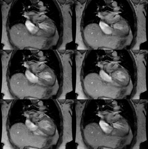
Asplenia (Ivemark's syndrome): Bilateral Right-sidedness, Right Isomerism, Heterotaxy syndrome with asplenia
Clinical:
In right isomerism, those structures which are normally seen on the right side of the body are seen on both sides (bilateral right-sidedness) [5]. Patients are generally identified in the newborn or infant period with severe cyanosis and survival beyond one year of age is seen in under 5% of cases (mortality of 80-90% in the first year of life [6]). Males are affected twice as commonly as females [5].Right isomerism is associated with asplenia which occurs in 42% of patients [7].Asplenia is characterized by:
1- Ambiguous abdominal situs- large transverse liver, midline stomach, and a common gastrointestinal mesentery with malrotation- however, abdominal situs ambiguous may not be evident on plain films in up to 50% of cases [2]. Other abnormalities include gallbladder agenesis, imperforate anus, horseshoe kidney, and posterior urethral valves [5].
2- Bilateral tri-lobed lungs and eparterial bronchi (mainstem bronchi pass over the pulmonary arteries and will be seen anterior to the bronchi on cross sectional images).
3- Splenic agenesis (Absence of the spleen also places these patients are increased risk of infection).
4- Persistent left SVC (bilateral SVC in 72% of cases).
5- Inapparent azygous vein.
6- Severe cardiac defects- Asplenia is almost always associated
(94-100%
of cases) with severe, complex, and inoperable cardiac
malformations.
Total anaomalous pulmonary venous drainage (seen in almost 50% of
cases), atrioventricular canal, transposition of the great
arteries, a
large
atrial defect or common atrium (75%), anomalies of pulmonary
venous
return
(62.5%), univentricular heart (37.5%), or pulmonary atresia. [6]
7- The presence of Howell-Jolly bodies (senescent RBC's on the peripheral smear due to absent spleen). Absent spleen also results in increased risk for life-threatening infection [5].
X-ray:
There is usually an associated complex congenital heart disease- single atrium with 2 right atrial appendages, pulmonary stenosis or atresia, and total anomalous pulmonary venous return.REFERENCES:
(1) J Thorac Imag, 1995, 10: p.43-57
(2) Radiology 1973; Freedom RM, et al. Radiographic viseral patterns in the asplenia syndrome. 106: 387-391
(3) Radiographics 1999; Applegate KE, et al. Situs revisited: Imaging of the heterotaxy syndrome. 19: 837-852
(4) Radiographics 2002; Fulcher AS, Turner MA. Abdominal manifestations of situs anomalies in adults. 22: 1439-1456
(5) AJR 2009; Ghosh S, et al. Anomalies of visceroatrial situs.
193:
1107-1117
(6) J Cardiovasc Comput Tomogr 2012; Balan A, et al Atrial
isomerism: a pictorial review. 6: 127-136
(7) J Cardiovasc Comput Tomogr 2013; Wolla CD, et al. Cardiovascular manifestations of heterotaxy and related situs abnormalities assessed with CT angiography. 7: 408-416


