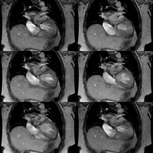
Aortic Interruption:
Clinical:
Interrupted aortic arch is defined by luminal discontinuity between the ascending and descending portions of the thoracic aorta- it may be complete or it may be spanned by an atretic fibrous band [5,6]. Complete interruption of the aortic arch (IAA) is a very rare anomaly - see in about 1% of infants with a critical congenital heart defect [5]. The clinical presentation of IAA is typically shock or severe heart failure in the first two weeks of life [6].IAA is associated with additional cardiovascular abnormalities in up to 98% of cases- most commonly a patent ductus arteriosus (about 97% of cases [2]) and usually a VSD (about 90% of cases [2]). A patent ductus is required to supply blood flow beyond the interrupted portion of the arch [2]. Other cardiac malformations are also common such as bicuspid aortic valve, muscular subaortic stenosis, atrial septal defects, and truncus arteriosus (about 10-20% of patients with truncus have an interrupted aortic arch).
Approximately 50% of IAA cases are associated with a chromosome 22q11.2 deletion (particularly in the presence of a right descending aorta) [2]. About 42% of patients with DiGeorge syndrome have an interrupted aortic arch [5]. The mortality rate is 80% in the first month and more than 90% at one year if untreated [2,3]. Therefore surgical correction is performed at the time of diagnosis [3]. Initial treatment consists of prostaglandin therapy to preserve patency of the ductus arteriosus [2]. Definitive treatment can be performed in a single or multistage process depending on the associated cardiac abnormalities [2,3]. Long term survival has improved and approaches 92% at one year after surgical repair [2], although, higher early post operative mortality rates have been reported (10-50%) [3].
The interruption is classified on the basis of the branches that originate from the proximal aorta and there are 3 types:
- Type A: In type A interruption there is no arch distal to the left subclavian artery and the descending aorta is a continuation of the patent ductus. This accounts for approximately 13-33% of IAA cases (it is the second most common form of IAA) [2,6]. The defect is likely the result of abnormal regression of the left 4th aortic arch after ascension of the left subclavian artery to its expected position [2]. Type A IAA is only very rarely found in patients with 22q11 deletion [6]. A Blalock-Park procedure is an anatomosis from the left subclavian artery to the descending aorta to bypass the interruption.
-
Type B: In type B, the interruption occurs between the left
carotid and
the left subclavian artery [6]. In this case, the left
subclavian originates
from the descending aorta. Type B interruption is the most
common type (about 66-84% of cases [2]). Type B IAA occurs when
the left 4th aortic arch regresses before normal ascension of
the left subclavian artery to its normal position [2]. A
chromosome 22q11.2 deletion is seen in up to 75% of patients
with type B IAA [2]. This chromosome deletion is associated with
thoracic midline malformations such as thymic hypoplasia and
conotruncal anomalies including subaortic stenosis, truncus
arteriosus, and tetralogy of Fallot [6]. There is an association
with DiGeorge's syndrome (which is congenital
absence of the thymus and parathyroid glands- between 10-42% of
patients with
this disorder have interruption of the aortic arch). A right
sided descending aorta with aortic interruption is almost always
associated with DiGeorge's syndrome [1]. Type B IAA is also
associated with bicuspid aortic valve [6].
- Type C: In type C, the interruption occurs between the innominate and left carotid arteries [2] (both the left carotid artery and left subclavian artery are connected to the descending aorta). This form is rare (less than 5% of cases) [2]. It occurs when the ventral portion of the left 3rd aortic arch and the left 4th aortic arch involute, and there is a persistent ductus caroticus ( a structure that normally regresses) [2].
REFERENCES:
(1) Radiographics 2003; Goo HW, et al. CT of congential heart
disease: normal anatomy and typical pathologic conditions. 23:
S147-S165
(2) AJR 2008; Dillman JR, et al. Interrupted aortic arch: spectrum of MRI findings. 190: 1467-1474
(3) Radiology 2008; Gaca AM, et al. Repair of congenital heart disease: a primer- part 2. 248: 44-60
(4) Radiographics 2010; Kimura-Hayama ET, et al. Uncommon congenital and acquired aortic diseases: role of multidetector CT angiography. 30: 79-98
(5) Radiographics 2010; Frank L, et al. Cardiovascular MR imaging
of conotruncal anomalies. 30: 1069-1094
(6) Radiographics 2017; Hanneman K, et al. Congenital variants and anomalies of the aortic arch. 37: 32-51


