Follicular Bronchiolitis/Follicular hyperplasia:
View cases of follicular bronchitis
Clinical:
Follicular bronchiolitis is rare benign polyclonal lymphoproliferative disorder characterized by lymphoid hyperplasia of bronchus-associated lymphoid tissue (BALT) in the airway wall [4,6]. The disorder is associated with underlying collagen vascular disorders such as juvenile rheumatoid arthritis and Sjogrens syndrome. It can also occur in association with chronic infection (bronchiectasis), congenital immune deficiency syndromes (common variable immune deficiency and Evan's syndrome), in AIDS, and in association with hypersensitivity disorders. Other less common associations include immunoglobulin A deficiency, Evans syndrome, Wiskott-Aldrich syndrome, and nylon and polyethylene exposures [7]. Treatment is directed at the underlying disease [7].Patients typically present with progressive shortness of breath and cough [3]. patients typically have a favorable prognosis, although patients under 30 years of age tend to have progressive disease [3]. Treatment is directed at the underlying disease [3,7]. If no identifiable cause is found (idiopathic), steroids or azothioprine, bronchodilator therapy, and macrolide antibiotics can be used for treatment [7]
Histologically there is proliferation of hyperplastic lymphoid follicles distributed the alveolar interstitium (resulting in expansion of the alveolar septa) and airway walls [1,6]. The disorder is more common in adults, but can occur at any age [3].
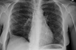
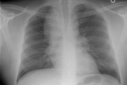
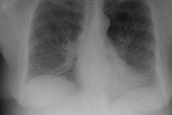
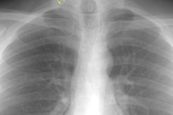
X-ray:
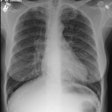
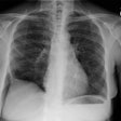
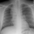
Plain films show a diffuse pattern of reticulonodular or miliary
opacities.
On HRCT the disorder is characterized by the presence of
scattered, small (generally about 3 mm in size), centrilobular
solid or ground-glass nodules [7]. The nodules can also be
diffusely distributed, or less commonly, peribronchial and
sub-pleural micronodules [7]. Larger nodules (3-12 mm) can be
found in about half of affected patients and ground-glass
opacities are seen in 75% of cases [3]. The centrilobular nodules
sometimes demonstrate a tree-in-bud configuration [6]. Air
trapping has been reported (due to lymphoid tissue narrowing of
the small airways) [1]. Thin walled cysts are a less common
feature [5].
REFERENCES:
(1) AJR 1999; Oh, Yu-Whan, et al. Follicular hyperplasia of
bronchus associated lymphoid tissue causing severe air trapping.
172: 745-747 (No abstract available)
(2) Radiology 1999; Howling SJ, et al. Follicular bronchitis: Thin-section CT and histologic findings. 212: 637-642
(3) Thorax 2001; Travis WD, Galvin JR. Non-neoplastic pulmonary lymphoid lesions. 56: 964-71. (No abstract available)
(4) AJR 2005; Pipavath SJ, et al. Radiologic and pathologic
features of bronchiolitis. 185: 354-363
(5) AJR 2011; Seaman DM, et al. Diffuse cystic lung disease at
high-resolution CT. 196: 1305-1311
(6) Radiographics 2016; Sirajuddin A, et al. Primary pulmonary
lymphoid lesions: radiologic and pathologic findings. 36: 53-70
(7) Radiographics 2017; Winningham PJ, et al. Bronchiolitis: a practical approach for the general radiologist. 37: 777-794
