Alveolar Microlithiasis:
View cases of alveolar microlithiasis
Clinical:
Alveolar
microlithiasis is autosomal recessive disorder
characterized by the development of sand-like calcifications
composed of calcium and phosphorus within the alveoli [5],
however, sporadic cases may occur [6]. The disease is caused
by autosomal recessive
inheritance of a mutation in the gene that produces the
protein that is the only known sodium-dependent phosphate
transporter protein in the lungs [5]. As a result, patients
are unable to transport phosphorus ions from the alveolar
space into type II pneumocytes
which leads to the development of calcospherites
in the alveolar space [5]. Identification of the SLC34A2 gene
mutation is diagnostic of PAM [6].
Patients
generally
present between the ages of 30 to 50 years, although the
condition has been identified in utero.
There is no sex predilection (although sporadic
cases are more common in males) [6]. There is a
positive family history in 50% of cases, especially siblings.
In the early phase of the disease, the lung tissue remains
normal, however, as the microliths expand, the alveolar walls
become compressed and damaged and eventually replaced by
fibrous tissue [6]. Over time (20-30 years), as the microliths
progress to involve all lobes bilaterally, the lungs increase
in weight and lose their collapsibility [6]. Most patients are
asymptomatic (70%), but DOE or clubbing may be seen (symptoms
typically do not develop until the 3rd or 4th decade of life
[6]). With advanced disease, patients may expectorate
microliths [6].
The disorder is slowly progressive and usually fatal due to
respiratory or cardiac failure. Patients have normal serum
calcium and phosphorus levels, and parathyroid hormone levels
are also normal [6]. On pulmonary function testing a decreased
residual volume or restrictive pattern are identified [6].
There is currently no effective treatment for the disorder
other than lung transplantation [5,6]. A differential
consideration includes talc granulomatosis secondary to IV
drug use.
X-ray:
CXR:
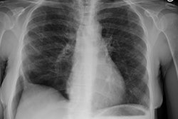
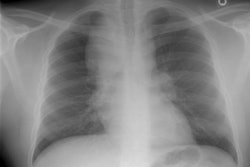
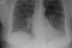
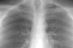
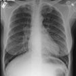
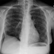
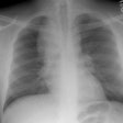
On plain film there is fine, sand-like micronodulation
(<1mm) involving both lungs diffusely (but more prominent
in the mid and lower lung zones [6]) and may appear confluent
in areas (producing ground glass opacity). There is a
predominantly symmetric middle and lower lobe involvement. The
heart borders and the diaphragm are usually obliterated. The
pattern has been described as ?sandstorm-like? [5]. A "black
pleural line" or zone of increased translucence between the
lung parenchyma and the ribs has been described and is due to
the presence of thin-walled subpleural
cysts [6]. Subpleural cyst rupture can lead to pneumothorax
[6]. With time, interstitial fibrosis, pulmonary hypertension,
and cor pulmonale
develop. The major finding on chest
radiographs in children are ground-glass opacifications.
Computed Tomography: On CT, there is a gradient in the distribution of the calcifications which tend to cluster in the posterior and inferior subpleural spaces, the anterior segments of the upper lobes, and along the bronchovascular bundles. Additionally, the medial portions of the lung appear to be more heavily involved than the peripheral portions [5]. Calcific polygonal lines can also be seen due to thickening of the interlobular septa producing a crazy paving pattern [6]. Calcified nodules larger than 1 mm (up to 5 mm) are visible on HRCT scans. Microliths with a diameter of less than 1 mm produce areas of ground-glass opacification, but can be discerned as discrete calcifications with appropriate windowing [5]. Numerous, thin walled sub-pleural cysts produce a dark pleural line and are indicative of early fibrosis.
Scintigraphy: There is intense pulmonary uptake of radiotracer on Tc-MDP bone scan [6].
REFERENCES:
(1) AJR 1997; Helbich TH, et al. Pulmonary alveolar microlithiasis in children: radiographic and high-resolution CT findings. Jan 168 (1): 63-65 (No abstract available)
(2) J Comput Assist Tomogr 1991; Pulmonary alveolar microlithiasis: CT findings. 15 (6): 938-42
(3) Pediatr Radiol 1987; Volle E, et al. Pulmonary alveolar microlithiasis in pediatric patients- review of the world literature and two new observations. 17: 439-442
(4) AJR Am J Roentgenol 1992; Korn MA, et al. Pulmonary alveolar microlithiasis: findings on high-resolution CT. 158(5): 981-982 (No abstract available)
(5) Radiographics 2011; Siddiqui NA, Fuhrman CR. Pulmonary
alveolar microlithiasis. 31:
585-590
(6) Radiographics 2016; Delic JA, et al. Pulmonary alveolar
microlithiasis. 36: 1334-1338
(7) Radiology 2021; Ufuk F. Pulmonary alveolar microlithiasis.
298: 567
