Endocardial Cushion Defects/Atrioventricular Septal Defect:
Clinical:
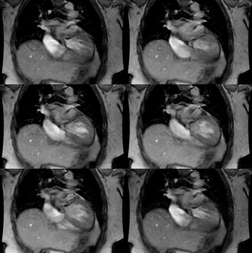
Endocardial cushion defects (or atrioventricular septal defects [4]) account for about 4% of all cases of congenital heart disease and represent a spectrum of anomalies that result from interruption of the normal development of the endocardial cushions during gestation [2]. Approximately 40% of patients with endocardial cushion defects have Down's syndrome. The lesion can also be found in patient with asplenia/polysplenia. The endocardial cushion normally forms the lower portion of the atrial septum, the upper portion of the ventricular septum, the septal leaflets of the mitral valve, and the tricuspid valve [2]. Sub-types of endocardial cushion defects include: (**Note- All have ostium primum ASD defect)1- Complete: Consists of an ostium primum ASD, a large posterior VSD, and a common atrioventricular valve to the RV & LV with 5 to 6 leaflets. As a result, there is free regurgitation from the ventricles into the atria at the AV valve level [3]. All four chambers of the heart communicate and there is both left-to-right and right-to-left shunting [4]. Patients present with CHF. The condition is associated with mesocardia/dextrocardia.
2- Intermediate
3- Partial: This is the most common form. Patient have two A-V valves and two A-V valve rings. There is an ostium primum ASD, but no VSD. These patient are usually not symptomatic in childhood. The mitral valve is tri-leaflet with a characteristic cleft in the anterior leaflet. The communication is between the RA-LA or RA-LV; occ. LV-RV.
X-ray:
On CXR there is shunt vascularity and LAE (due to mitral valve incompetence), coupled with right heart enlargement. A "gooseneck sign" is characteristic on angiography- it is caused by a deficiency of both the conus and sinus portions of the interventricular septum, with narrowing of the left ventricular outflow tract [2]. The concavity of the interventricular septum below the mitral valve, along with elongation and narrowing of the LV outflow tract, produces a characteristic shape "gooseneck" shape on the AP projection at angiography [2].
Endocardial cushion defect: 22 d patient presented with SOB. Note cardiac enlargement and marked increased vascularity in the lungs (shunt vascularity). Cardiac echo revealed an endocardial cushion defect. The patient did not have Down's syndrome. (Click image to enlarge) |
|
|
REFERENCES:
(1) Pediatric Clinics of North America 1999; Driscoll DJ. Left-to-right
shunt lesions. 46(2): 355-368
(2) Radiographics 2007; Ferguson EC, et al. Classic imaging signs of congenital cardiovascular abnormalities. 27: 1323-1334
(3) Radiographics 2002; Barboza JM, et al. Prenatal diagnosis of congenital cardiac anomalies: a practical approach using two basic views. 22: 1125-1138
(4) Radiographics 2003; Wang ZJ, et al. Cardiovascular shunts: MR imaging evaluation. 23: S181-S194


