Pulmonary alveolar proteinosis: high-resolution CT, chest radiographic, and functional correlations.
Lee KN, Levin DL, Webb WR, Chen D, Storto ML, Golden JA
Department of Radiology, University of California, San Francisco, USA.
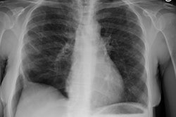
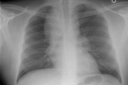
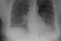
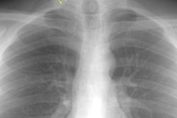
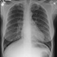
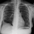
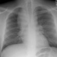
STUDY OBJECTIVE: To determine whether a correlation exists between pulmonary function and both frontal chest radiographs and high-resolution chest CT findings in patients with pulmonary alveolar proteinosis (PAP). DESIGN: Retrospective review of radiographic and clinical data. SETTING: Tertiary referral hospital. PATIENTS: Seven patients with PAP were studied on 25 occasions using high-resolution chest CT (n=21), frontal chest radiographs (n=19), and pulmonary function tests (PFTs) (n=25). MEASUREMENTS AND RESULTS: Visual estimates of the extent, degree, and overall severity of parenchymal abnormalities were determined for plain radiographs and high-resolution chest CT, and were correlated with PFTs. With high-resolution CT, the extent and severity of ground-glass opacity correlated significantly with the presence of a restrictive ventilatory defect, reduced diffusing capacity, and hypoxemia. Chest radiographic findings also correlated significantly with restrictive ventilatory defect, diffusing capacity, and hypoxemia. CONCLUSION: In patients with PAP, although high-resolution CT correlates more closely with pulmonary function, plain radiographs should be sufficient for follow-up.
